

Primary systemic amyloidosis: imaging interpretation of this complex multisystemic disease
- PMID: 30460006
- PMCID: PMC6243301
- DOI: 10.1259/bjrcr.20150171
Primary systemic amyloidosis: imaging interpretation of this complex multisystemic disease
Abstract
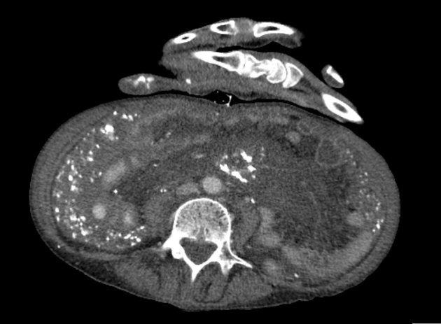
This report highlights the diagnostic complexities involved in the case of a 63-year-old female who presented with a non-productive cough and shortness of breath on exertion. Initial chest radiograph demonstrated generalized abnormal interstitial lung markings with thickened peripheral septal lines. Further characterization was sought by CT scan of the chest, and given the possibility of lymphangitic carcinomatosis, a CT scan of the abdomen and pelvis was also performed. The CT scan findings revealed septal line thickening, abnormal omental soft tissue with calcified deposits and wall thickening of the stomach and proximal duodenum. A preliminary differential diagnosis of peritoneal carcinomatosis was made, but cancer markers were equivocal. A CT-guided biopsy of the "omental cake" was non-diagnostic, hence formal biopsy via laparoscopy was undertaken. While awaiting the results, the patient was readmitted with acute haematemesis. Gastric and duodenal biopsies from the endoscopic assessment were positive for Congo red stain and birefringent under polarizsed light, which was consistent with amyloidosis. Histology from the omental biopsies and additional haematological tests concurred. The patient was diagnosed with advanced systemic amyloid light-chain amyloidosis comprising diffuse pulmonary amyloidosis, calcified omental soft tissue deposits, and extensive soft tissue amyloid with cardiac and gastrointestinal involvement. We discuss the spectrum of differential diagnoses posed by the imaging findings and the difficulties faced in interpreting this complex case of systemic amyloidosis.
Figures


References
-
- Vicens RA, Patnana M, Le O, Bhosale PR, Sagebiel TL, Menias CO, et al. . Multimodality imaging of common and uncommon peritoneal diseases: a review for radiologists. Abdom Imaging 2015; 40: 436–56. - PubMed
-
- Agarwal A, Yeh BM, Breiman RS, Qayyum A, Coakley FV. Peritoneal calcification: causes and distinguishing features on CT. AJR Am J Roentgenol 2004; 182: 441–5. - PubMed
-
- National Amyloidosis Centre. UCL Division of Medicine; c1999-2015 [updated 9 January 2015; cited 8 February 2015]. Available from: http://www.ucl.ac.uk/amyloidosis/nac
-
- Sayed RH, Hawkins PN, Lachmann HJ. Emerging treatments for amyloidosis. Kidney Int 2015; 87: 516–26http://www.nature.com/ki/journal/vaop/ncurrent/full/ki2014368a.html. - PubMed
Publication types
LinkOut - more resources
Full Text Sources
