CT and MR of recurrent primary cutaneous adenoid cystic carcinoma with multiple metastases
- PMID: 30460012
- PMCID: PMC6243298
- DOI: 10.1259/bjrcr.20150243
CT and MR of recurrent primary cutaneous adenoid cystic carcinoma with multiple metastases
Abstract
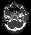
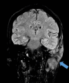
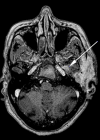
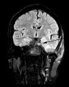
Primary cutaneous adenoid cystic carcinoma is a rare slow-growing neoplasm, with limited literature reporting the involvement of the scalp. It has a tendency to recur locally; however, lymph node, distant pulmonary and bony metastases are exceptionally rare. We highlight the case of a 65-year-old female with primary cutaneous adenoid cystic carcinoma with distant pulmonary and bony metastases and the importance of imaging in diagnosing distant metastasis and perineural spread.
Figures


Similar articles
-
Primary cutaneous adenoid cystic carcinoma with brain metastases: case report and literature review.J Cutan Pathol. 2016 Feb;43(2):137-41. doi: 10.1111/cup.12563. Epub 2015 Sep 11. J Cutan Pathol. 2016. PMID: 26238986 Review.
-
Primary cutaneous adenoid cystic carcinoma with lymph node metastasis.Am J Dermatopathol. 1998 Dec;20(6):571-7. doi: 10.1097/00000372-199812000-00005. Am J Dermatopathol. 1998. PMID: 9855352 Review.
-
Lung metastases from cutaneous adenoid cystic carcinoma 23 years after initial treatment.Respir Med Case Rep. 2017 Apr 19;21:121-123. doi: 10.1016/j.rmcr.2017.04.015. eCollection 2017. Respir Med Case Rep. 2017. PMID: 28462081 Free PMC article.
-
Primary cutaneous adenoid cystic carcinoma with regional lymph node metastasis.J Laryngol Otol. 2001 Aug;115(8):673-5. doi: 10.1258/0022215011908595. J Laryngol Otol. 2001. PMID: 11535157 Review.
-
Intraoral adenoid cystic carcinoma: is the presence of perineural invasion associated with the size of the primary tumour, local extension, surgical margins, distant metastases, and outcome?Br J Oral Maxillofac Surg. 2014 Mar;52(3):214-8. doi: 10.1016/j.bjoms.2013.11.009. Epub 2013 Dec 8. Br J Oral Maxillofac Surg. 2014. PMID: 24325947
Cited by
-
Cutaneous Adenoid Cystic Carcinoma Presenting as Proliferative Nodules - A Case Report.Indian Dermatol Online J. 2024 Oct 11;15(6):1033-1035. doi: 10.4103/idoj.idoj_743_23. eCollection 2024 Nov-Dec. Indian Dermatol Online J. 2024. PMID: 39640456 Free PMC article. No abstract available.
References
-
- Khan AJ, DiGiovanna MP, Ross DA, Sasaki CT, Carter D, Son YH, et al. . Adenoid cystic carcinoma: A retrospective clinical review. Int J C 2001; 96: 149–58. - PubMed
-
- Fueston JC, Gloster HM, Mutasim DF. Primary cutaneous adenoid cystic carcinoma: A case report and literature review. Cutis 2006; 77: 157–60. - PubMed
-
- Boggio R. Adenoid cystic carcinoma of scalp. [Letter.]. Arch Dermatol 1975; 111: 793–4. - PubMed
-
- Naylor E, Sarkar P, Perlis CS, Giri D, Gnepp DR, Robinson-Bostom L. Primary cutaneous adenoid cystic carcinoma. J Am Acad Dermatol 2008; 58: 636–41. - PubMed
Publication types
LinkOut - more resources
Full Text Sources

