

An unusual case of intercostal muscle flap ossification mimicking an intrathoracic rib
- PMID: 30460028
- PMCID: PMC6243329
- DOI: 10.1259/bjrcr.20150469
An unusual case of intercostal muscle flap ossification mimicking an intrathoracic rib
Abstract
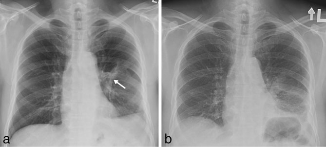
We present a unique case of intercostal muscle flap (ICMF) ossification mimicking an intrathoracic rib diagnosed 3 years after oesophageal perforation repair. A 58-year-old male presented with complaints of mild chest discomfort. Three years ago he had undergone left thoracotomy and primary repair of post-emetic oesophageal perforation. An ICMF had been used to strengthen the repair. Chest X-ray identified a linear calcific density within the left hemithorax. Subsequent thoracic CT characterized the anomaly as ossification of the ICMF. The lesion had the appearance of a well-differentiated intrathoracic rib coursing through the left lower lobe. We discuss the typical appearances of ossified ICMFs and the potential complications resulting from this ossification.
Figures


References
-
- Prommegger R, Salzer GM. Heterotopic ossification in pedicled intercostal muscle flaps causing clinical problems. J Thorac Cardiovasc Surg 1998; 115: 466–7. - PubMed
-
- Kwek BH, Wain JC, Aquino SL. The radiologic appearance of intercostal muscle flap. Ann Thorac Surg 2004; 78: 432–5. - PubMed
-
- Deeb ME, Sterman DH, Shrager JB, Kaiser LR. Bronchial anastomotic stricture caused by ossification of an intercostal muscle flap. Ann Thorac Surg 2001; 71: 1700–2. - PubMed
-
- Cerfolio RJ, Bryant AS, Yamamuro M. Intercostal muscle flap to buttress the bronchus at risk and the thoracic esophageal-gastric anastomosis. Ann Thorac Surg 2005; 80: 1017–20. - PubMed
-
- Alexander PV, Hollands M, O'Rourke IC, Tait N. Intercostal pedicle flap for thoracic oesophageal perforations. ANZ J Surg 1997; 67: 133–5. - PubMed
Publication types
LinkOut - more resources
Full Text Sources
