

Case Reports
doi: 10.1259/bjrcr.20150507.
eCollection 2016.
Pancreatic lipoma: a pancreatic incidentaloma; diagnosis with ultrasound, computed tomography and magnetic resonance imaging
Affiliations
- PMID: 30460031
- PMCID: PMC6243312
- DOI: 10.1259/bjrcr.20150507
Item in Clipboard
Case Reports
Pancreatic lipoma: a pancreatic incidentaloma; diagnosis with ultrasound, computed tomography and magnetic resonance imaging
BJR Case Rep.
.
Abstract
Pancreatic lipomas are rare. We present a case of incidentally discovered pancreatic lipoma in a 45-year-old female suffering from metastatic ovarian carcinoma who was referred to radiology for follow-up imaging. Fat-containing tumours originating from the pancreas are very rare. Most lipomasshow characteristic features on imaging that allow their differentiation. In most cases, accurate diagnosis is attained without any histopathological confirmation. We present the imaging features of pancreatic lipoma on ultrasound, CT scan and MRI, the differential diagnosis and a brief review of the literature.
Figures
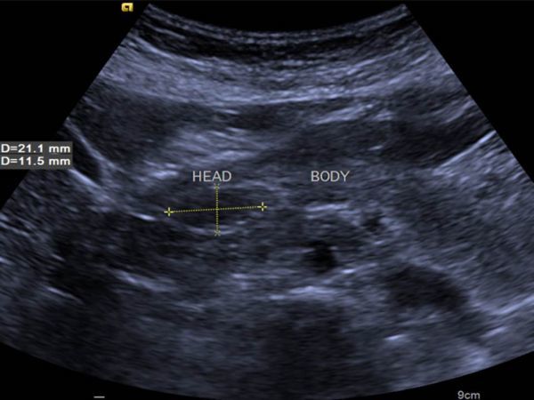
Transverse grayscale (B-mode) ultrasound image showing hypo- to isoechoic lesion on the head of the pancreas.
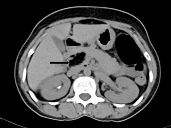
Non-contrast CT scan shows a well-circumscribed focal lesion (arrow) on the pancreatic head, measuring 35 x 35 x 19 mm and with a density of −106 Hounsfield units, consistent with fatty tissue.
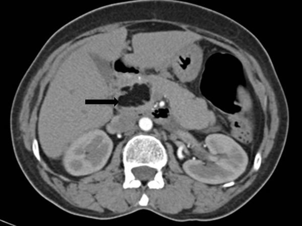
Contrast-enhanced CT scan shows a homogeneous focal mass (arrow) on the pancreatic head. The mass was isodense with fatty tissue and interlobular septa, and without central or peripheral contrast enhancement (arrow).
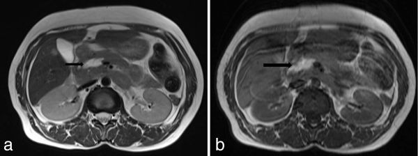
(a) Axial T2 and (b) T1 weighted MRI showing a hyperintense lobulated mass (arrows) on the head of the pancreas.
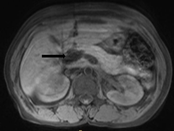
Axial T1 weighted fat-suppressed sequence showing suppression of T1 hyperintensity (black arrow) within the lesion, suggesting a lesion of fatty nature.
Similar articles
-
Pancreatic Lipoma: A Pancreatic Incidentaloma Diagnosis with Computed Tomography.Eur J Case Rep Intern Med. 2021 Mar 23;8(3):002252. doi: 10.12890/2021_002252. eCollection 2021. Eur J Case Rep Intern Med. 2021. PMID: 33869089 Free PMC article.
-
Large pancreatic lipoma in a 69-year-old diabetic woman: diagnostic considerations.Prz Gastroenterol. 2014;9(3):168-71. doi: 10.5114/pg.2014.43579. Epub 2014 Jun 26. Prz Gastroenterol. 2014. PMID: 25097715 Free PMC article.
-
Lipoma of the pancreas, a case report and a review of the literature.World J Radiol. 2011 Oct 28;3(10):246-8. doi: 10.4329/wjr.v3.i10.246. World J Radiol. 2011. PMID: 22229078 Free PMC article.
-
Small pancreatic lipoma: case report and literature review.Hepatogastroenterology. 2007 Jul-Aug;54(77):1582-4. Hepatogastroenterology. 2007. PMID: 17708305 Review.
-
Fat containing unusual tumor of the pancreas.Eur Radiol. 2002 Apr;12(4):770-3. doi: 10.1007/s003300101032. Epub 2001 Sep 6. Eur Radiol. 2002. PMID: 11960224 Review.
Cited by
-
Pancreatic ductal adenocarcinoma arising from the pancreatic parenchyma compressed by a huge pancreatic lipoma: a case report.Surg Case Rep. 2023 Aug 1;9(1):136. doi: 10.1186/s40792-023-01720-w. Surg Case Rep. 2023. PMID: 37526778 Free PMC article.
-
Pancreatic Lipoma: A Pancreatic Incidentaloma Diagnosis with Computed Tomography.Eur J Case Rep Intern Med. 2021 Mar 23;8(3):002252. doi: 10.12890/2021_002252. eCollection 2021. Eur J Case Rep Intern Med. 2021. PMID: 33869089 Free PMC article.
-
Pancreatic changes with lifestyle and age: What is normal and what is concerning?Endosc Ultrasound. 2023 Mar-Apr;12(2):213-227. doi: 10.4103/EUS-D-22-00162. Endosc Ultrasound. 2023. PMID: 37148135 Free PMC article. Review.
-
Concurrent Pancreatic and Sartorius Muscle Lipomas: A Case Report.Clin Case Rep. 2025 Apr 25;13(5):e70350. doi: 10.1002/ccr3.70350. eCollection 2025 May. Clin Case Rep. 2025. PMID: 40291570 Free PMC article.
-
Unveiling a rare surgical phenomenon: Symptomatic pancreatic lipoma's diagnosis and management.Int J Surg Case Rep. 2025 Sep;134:111709. doi: 10.1016/j.ijscr.2025.111709. Epub 2025 Jul 23. Int J Surg Case Rep. 2025. PMID: 40784103 Free PMC article.
References
-
- Ferrozzi F, Cusmano F, Zuccoli G, Tognini G, Bassi S, Gabrielli M. Mesenchymal tumors of the pancreas: computed tomography patterns. Radiol Med 1999;98: 295–9. - PubMed
-
- Itai Y, Saida Y, Kurosaki Y, Kurosaki A, Fujimoto T. Focal fatty masses of the pancreas. Acta Radiol 1995;36: 178–81. - PubMed
-
- De Jong SA, Pickleman J, Rainsford K. Nonductal tumors of the pancreas. The importance of laparotomy. Arch Surg 1993;128: 730–4. - PubMed
-
- Bigard MA, Boissel P, Regent D, Froment N. First case in the literature. Gastroentérologie Clin Biol 1989;13: 505–7. - PubMed
-
- Raut CP, Fernandez-del Castillo C. Giant lipoma of the pancreas: case report and review of lipomatous lesions of the pancreas. Pancreas 2003;26: 97–9. - PubMed
Publication types
LinkOut - more resources
Full Text Sources
