. 2017 Jan 15;21(1):10-16.
doi: 10.1080/19768354.2016.1276025. eCollection 2017.

Distinct regulations of ARF1 by two Aplysia Sec7 isoforms

Affiliations

Distinct regulations of ARF1 by two Aplysia Sec7 isoforms

Yong-Woo Jun et al. Anim Cells Syst (Seoul). .

Abstract

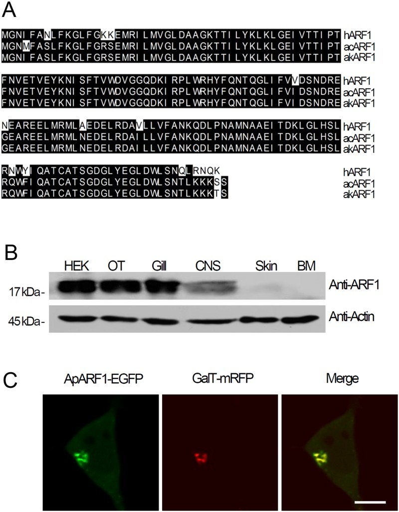
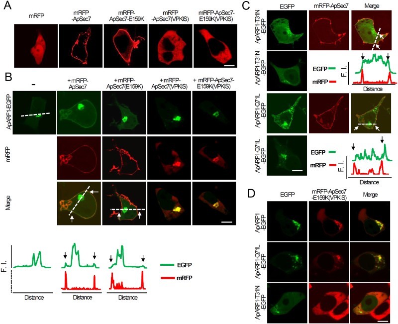
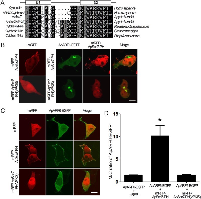
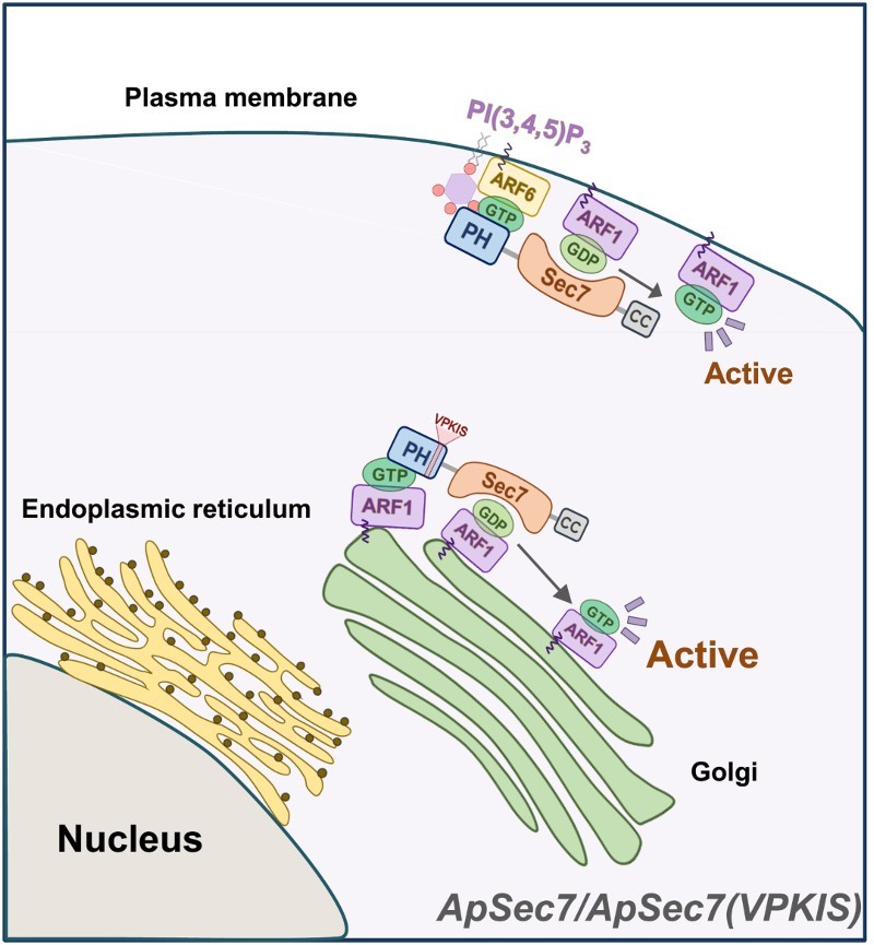
Sec7 protein is a guanine nucleotide exchange factor in the ADP-ribosylation factor (ARF) family of small GTP-binding proteins. Aplysia Sec7 proteins (ApSec7s) play many roles in neurite outgrowth and synaptic facilitation in Aplysia neurons. However, the binding property of Aplysia ARF1 by ApSec7 isoforms has not been examined. In this study, we found that the cloned Aplysia ARF1 (ApARF1) protein only localized to the Golgi complex when it was expressed alone in HEK293T cells; however, if ApARF1 was co-expressed with plasma membrane-targeted ApSec7, it localized to both the plasma membrane and the Golgi complex via association with the Sec7 domain of ApSec7. Moreover, in HEK293T cells expressing both ApARF1 and another Sec7 isoform, ApSec7(VPKIS), the pleckstrin homology domain of ApSec7(VPKIS) associated with ApARF1, resulting in its localization to the Golgi complex. Overall, we propose a model in which ApSec7(VPKIS) activates ApARF1 in the Golgi complex, while ApSec7 recruits ApARF1 to the plasma membrane where it activates ApARF1/6 downstream signaling.

Keywords: ARF1; ARF6; ApSec7; ApSec7(VPKIS); Aplysia.


Figures

Figure 1.
Figure 1.
Cloning and tissue expression of ApARF1 from Aplysia kurodai. (a) Multiple sequence alignment of ARF1 from Aplysia kurodai (akARF1), Aplysia californica (acARF1), and human (hARF1). ARF1, ADP-ribosylation factor 1: Human, Homo sapiens; Aplysia, A. kurodai (NCBI Accession number: KU724187). (b) Western blotting of ApARF1 in lysates from various Aplysia tissues and HEK293T cells. HEK, HEK293T cells; OT, ovotestis; Gill, gill; CNS, central nervous system, including pleural, pedal, central, and abdominal ganglion; BM, buccal muscle. (c) Cellular localization of ApARF1-EGFP in HEK293T cells; GalT-mRFP was used as a Golgi marker. (d–e) Effects of ApSec7 expression on the cellular localization of ApARF1 in HEK293T cells. Scale bar, 20 μm.
Figure 2.
Figure 2.
Effects of ApSec7 expression on the cellular localization of ApARF1 in HEK293T cells. (a–b) Effects of ApSec7 expression on the cellular localization of ApARF1 in HEK293T cells. (a) mRFP-ApSec7 and mRFP-ApSec7-E159K were localized to the plasma membrane, whereas mRFP, mRFP-ApSec7(VPKIS), and mRFP-ApSec7-E159K(VPKIS) were localized to the cytosol in HEK293T cells. Scale bar, 20 μm. (b) Cellular localization of mRFP-ApSec7 and ApARF1-EGFP in HEK293T cells. ApARF1-EGFP was localized to the plasma membrane only in mRFP-ApSec7- and ApARF1-EGFP-expressing cells. In contrast, mRFP-ApSec7(VPKIS) and mRFP-ApSec7-E159K(VPKIS) were localized to the Golgi complex in mRFP-ApSec7(VPKIS)- and ApARF1-EGFP-expressing cells or in mRFP-ApSec7-E159K(VPKIS)- and ApARF1-EGFP-expressing cells. The white dashed lines in the confocal fluorescence images indicate the paths along which the fluorescence intensities (F.I.) of the corresponding images were plotted. Black (traces) or white arrows (cell images) indicate the corresponding positions of the plasma membrane targeting mRFP signaling. Scale bar, 20 μm. (c) Cellular localization of ApARF1-T31N-EGFP or ApARF1-Q71L-EGFP in the presence of mRFP-ApSec7. The white dashed lines in the confocal fluorescence images indicate the paths along which the fluorescence intensities (F.I.) of the corresponding images were plotted. Black (traces) or white arrows (cell images) indicate the corresponding positions of the plasma membrane targeting mRFP signaling. Scale bar, 20 μm. (d) Cellular localization of ApARF1-EGFP, ApARF1-T31N-EGFP, or ApARF1-Q71L-EGFP in the presence of mRFP-ApSec7-E159K(VPKIS). Scale bar, 20 μm.
Figure 3.
Figure 3.
Binding of the PH domain of ApSec7(VPKIS) to ARF1. (a) Multiple sequence alignment of the PH domain of cytohesin family members of various species: cytohesin4 and ARNO/cytohesin2 from Homo sapiens, ApSec7 and ApSec7(VPKIS) from Aplysia kurodai, and cytohesin1-like proteins from Parasteatoda tepidariorum (NCBI Accession number: XP_015921179), Crassostrea gigas (NCBI Accession number: XP_011436446), and Priapulus caudatus (NCBI Accession number: XP_014669585). (b) Cellular localization of mRFP-ApSec7-PH or mRFP-ApSec7-PH(VPKIS) in the presence of ApARF1-EGFP. In the presence of ApARF1-EGFP, mRFP-ApSec7-PH(VPKIS) but not mRFP-ApSec7-PH was localized to the Golgi complex. Scale bar, 20 μm. (c–d) Enhancement of the plasma membrane localization of ApARF6-EGFP in the presence of mRFP-ApSec7-PH but not in the presence of mRFP-ApSec7-PH(VPKIS). Cellular localization of ApARF6-EGFP in the presence of mRFP, mRFP-ApSec7-PH, or mRFP-ApSec7-PH(VPKIS) in HEK293T cells. (c) Quantification of the ratio of the fluorescence intensity between the plasma membrane and cytosol localization (M/C ratio) of ApARF6-EGFP in HEK293T cells. *, p < .001 compared to that of all other groups, one-way ANOVA; F = 14.7, Tukey’s post-hoc test. Scale bar, 20 μm.
Figure 4.
Figure 4.
Schematic diagram of the molecular mechanism underlying the distinct functions of ApSec7 and ApSec7(VPKIS) in cells. The PH domain of ApSec7 was bound to PI(3,4,5)P3 and active ARF6, leading to plasma membrane localization, while the PH domain of ApSec7(VPKIS) was only weakly bound to active ARF1, leading to Golgi targeting. ApSec7(VPKIS) in the Golgi complex activated ApARF1 at the Golgi complex. In contrast, ApSec7 recruited ApARF1 to the plasma membrane via the Sec7 domain and activated ApARF1 at the plasma membrane.

Similar articles

References

    1. Casanova JE. 2007. Regulation of arf activation: the Sec7 family of guanine nucleotide exchange factors. Traffic. 8:1476–1485. doi: 10.1111/j.1600-0854.2007.00634.x - DOI - PubMed
    1. Cohen LA, Honda A, Varnai P, Brown FD, Balla T, Donaldson JG. 2007. Active Arf6 recruits ARNO/cytohesin GEFs to the PM by binding their PH domains. Mol Biol Cell. 18:2244–2253. doi: 10.1091/mbc.E06-11-0998 - DOI - PMC - PubMed
    1. Donaldson JG, Jackson CL. 2011. ARF family G proteins and their regulators: roles in membrane transport, development and disease. Nat Rev Mol Cell Biol. 12:362–375. doi: 10.1038/nrm3117 - DOI - PMC - PubMed
    1. D’Souza-Schorey C, Chavrier P.. 2006. ARF proteins: roles in membrane traffic and beyond. Nat Rev Mol Cell Biol. 7:347–358. doi: 10.1038/nrm1910 - DOI - PubMed
    1. Hernandez-Deviez DJ, Roth MG, Casanova JE, Wilson JM. 2004. ARNO and ARF6 regulate axonal elongation and branching through downstream activation of phosphatidylinositol 4-phosphate 5-kinase alpha. Mol Biol Cell. 15:111–120. doi: 10.1091/mbc.E03-06-0410 - DOI - PMC - PubMed

LinkOut - more resources