Review
. 2018 Aug 12;22(4):213-218.
doi: 10.1080/19768354.2018.1508498. eCollection 2018.

Exploring glia to better understand Alzheimer's disease

Affiliations
Review

Exploring glia to better understand Alzheimer's disease

Yoo Sung Kim et al. Anim Cells Syst (Seoul). .

Abstract

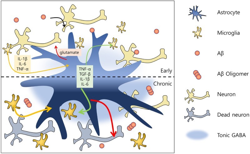
The amyloid-β (Aβ) hypothesis has been the leading explanation for the pathogenesis of Alzheimer's disease (AD). The most common traits of AD are cognitive impairments and memory loss, which are associated with the accumulation of Aβ. Aβ aggregates activate glial cells, which in turn remove Aβ. Because microglia act as immune cells in the brain, most glia-related studies of AD have focused primarily on this cell type. However, astrocytes, another type of glial cell, also participate in the brain immune system, synaptic formation, brain homeostasis, and various other brain functions. Accordingly, many studies on the underlying mechanisms of AD have investigated not only neurons but also glial cells. Although these studies suggest that microglia and astrocytes are effective targets for AD therapeutics, other recent studies have raised questions regarding whether microglial cells and/or astrocytes serve a neuroprotective or neurotoxic function in AD. To gain a better understanding of the mechanisms of AD and identify novel targets for AD treatment, in this review, we consider the role of both microglia and astrocytes in AD.

Keywords: Alzheimer’s disease; astrocytes; microglia; neuroglia.


Figures

Figure 1.
Figure 1.
Representative schematic of function of glial cells during the progression of AD. In the early stage of Alzheimer’s disease, microglial cells are activated by amyloid beta proteins and release cytokines and chemokines (e.g. IL-1β, IL-6, TNF-α). Reactive microglia then activate astrocytes, which release TNF-α, TGF-β, IL-1β and IL-6, as well as certain neurotransmitters (e.g. glutamate) that reciprocally influence microglia. The communication of glial cells and neurons with Aβ induces a chronic disease stage. In this phase, microglia and astrocytes positively feedback on each other, and are thus continuously activated. Excessive amounts of glutamate then become sufficiently neurotoxic to kill the neurons. At this neuronal death stage, increased tonic release of GABA by reactive astrocytes is detected.

Similar articles

Cited by

References

    1. Abramov AY, Duchen MR.. 2005. The role of an astrocytic NADPH oxidase in the neurotoxicity of amyloid beta peptides. Philos Trans R Soc Lond B Biol Sci. 360:2309–2314. doi: 10.1098/rstb.2005.1766 - DOI - PMC - PubMed
    1. Bahmanyar S, Higgins GA, Goldgaber D, Lewis DA, Morrison JH, Wilson MC, Shankar SK, Gajdusek DC.. 1987. Localization of amyloid beta protein messenger RNA in brains from patients with Alzheimer's disease. Science. 237(4810):77–80. doi: 10.1126/science.3299701 - DOI - PubMed
    1. Bezzi P, Domercq M, Brambilla L, Galli R, Schols D, De Clercq E, Vescovi A, Bagetta G, Kollias G, Meldolesi J, et al. . 2001. CXCR4-activated astrocyte glutamate release via TNFα: amplification by microglia triggers neurotoxicity. Nat Neurosci. 4:702–710. doi: 10.1038/89490 - DOI - PubMed
    1. Block ML, Zecca L, Hong JS.. 2007. Microglia-mediated neurotoxicity: uncovering the molecular mechanisms. Nat Rev Neurosci. 8:57–69. doi: 10.1038/nrn2038 - DOI - PubMed
    1. Cairns NJ, Atkinson PF, Hanger DP, Anderton BH, Daniel SE, Lantos PL.. 1997. Tau protein in the glial cytoplasmic inclusions of multiple system atrophy can be distinguished from abnormal tau in Alzheimer’s disease. Neurosci Lett. 230:49–52. doi: 10.1016/S0304-3940(97)00474-6 - DOI - PubMed

LinkOut - more resources