Review
. 2017;9(1):1.
doi: 10.1007/s40820-016-0103-7. Epub 2016 Aug 31.

Fabrication and Applications of Micro/Nanostructured Devices for Tissue Engineering

Affiliations
Review

Fabrication and Applications of Micro/Nanostructured Devices for Tissue Engineering

Tania Limongi et al. Nanomicro Lett. 2017.

Abstract

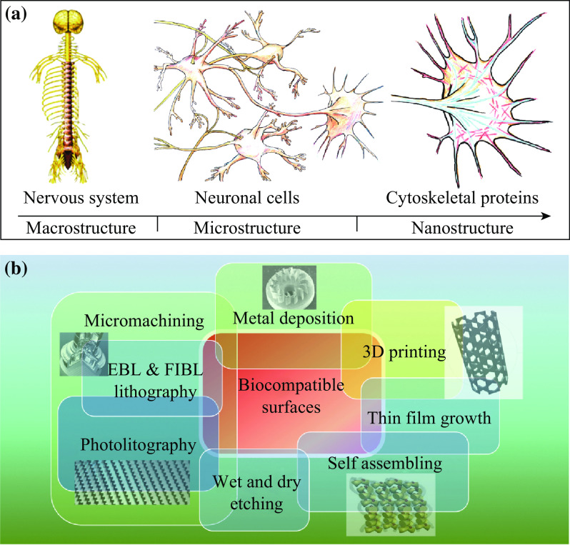
Nanotechnology allows the realization of new materials and devices with basic structural unit in the range of 1-100 nm and characterized by gaining control at the atomic, molecular, and supramolecular level. Reducing the dimensions of a material into the nanoscale range usually results in the change of its physiochemical properties such as reactivity, crystallinity, and solubility. This review treats the convergence of last research news at the interface of nanostructured biomaterials and tissue engineering for emerging biomedical technologies such as scaffolding and tissue regeneration. The present review is organized into three main sections. The introduction concerns an overview of the increasing utility of nanostructured materials in the field of tissue engineering. It elucidates how nanotechnology, by working in the submicron length scale, assures the realization of a biocompatible interface that is able to reproduce the physiological cell-matrix interaction. The second, more technical section, concerns the design and fabrication of biocompatible surface characterized by micro- and submicroscale features, using microfabrication, nanolithography, and miscellaneous nanolithographic techniques. In the last part, we review the ongoing tissue engineering application of nanostructured materials and scaffolds in different fields such as neurology, cardiology, orthopedics, and skin tissue regeneration.

Keywords: Device; Microfabrication; Nanofabrication; Nanomaterials; Nanostructures; Tissue engineering.


Figures

Fig. 1
Fig. 1
a Micro- and nanostructure of central and peripheral nervous systems. b The principal micro- and nanofabrication technologies for TE applications
Fig. 2
Fig. 2
Schematization of the fabrication process for 3D PCL pillared scaffolds using a hot press (on the left). a Silicon master production. b Micromolding melting step. c Micromolding pressing step. d Final structure obtained after solidification and detachment. Figure adapted from [17]
Fig. 3
Fig. 3
Nano-textured PCL film realized through a single-step plasma etching process. a The Silicon wafer acts as a support. b Embedding in cell culture medium. c Microfilm peeling-off for “free-standing” use. d AFM images of the nanostructured PCL surface. Figure adapted from [16]
Fig. 4
Fig. 4
a SEM micrograph showing a flat glial cell monolayer suspended between adjacent nanostructured pillars. b Low magnification of neuronal somas and its processes. c Neuronal projections densely wrap the pillar nanopatterned sidewall. Figure adapted from [15]
Fig. 5
Fig. 5
SEM images of primary hippocampal cultures plated on nanopatterned PCL substrates. a Neurons resulted healthy, as indicated by the smooth surface of cell bodies (asterisk), b Dense network of neurites (arrows), which grew in tight adhesion with the substrate. Figure adapted from [16]
Fig. 6
Fig. 6
Confocal images of primary hippocampal cultures plated on nanopatterned PCL substrates at two magnifications (upper and lower rows). Neuronal class III β-tubulin/synapsin I (a and a′), class III β-tubulin/neural cell adhesion molecule (b and b′), and class III β-tubulin/phosphorylated neurofilament proteins (c and c′). Figure adapted from [16]
Fig. 7
Fig. 7
SEM images of NIH/3T3 cells suspended on biocompatible PCL nanostructured micropillars. a Fibroblasts within 24 h produced filopodia sensing the microstructured biopolymer, and b thicker pseudopodia-like processes appeared to use pillars as stepping stones. Figure adapted from [18]

References

    1. Hwang J, Jeong Y, Park JM, Lee KH, Hong JW, Choi J. Biomimetics: forecasting the future of science, engineering, and medicine. Int. J. Nanomed. 2015;8(10):5701–5713. - PMC - PubMed
    1. Webber MJ, Appel EA, Meijer EW, Langer R. Supramolecular biomaterials. Nat. Mater. 2015;15(1):13–26. doi: 10.1038/nmat4474. - DOI - PubMed
    1. Luu TU, Gott SC, Woo BW, Rao MP, Liu WF. Micro- and nanopatterned topographical cues for regulating macrophage cell shape and phenotype. ACS Appl. Mater. Interfaces. 2015;7(51):28665–28672. doi: 10.1021/acsami.5b10589. - DOI - PMC - PubMed
    1. Mauro N, Manfredi A, Ranucci E, Procacci P, Laus M, Antonioli D, Mantovani C, Magnaghi V, Ferruti P. Degradable poly(amidoamine) hydrogels as scaffolds for in vitro culturing of peripheral nervous system cells. Macromol. Biosci. 2013;13(3):332–347. doi: 10.1002/mabi.201200354. - DOI - PubMed
    1. Zhu W, O’Brien C, O’Brien JR, Zhang LG. 3D nano/microfabrication techniques and nanobiomaterials for neural tissue regeneration. Nanomedicine. 2014;9(6):859–875. doi: 10.2217/nnm.14.36. - DOI - PubMed