. 2018 Dec;56(1):440-449.
doi: 10.1080/13880209.2018.1502326.

Resveratrol mitigates the oxidative stress mediated by hypoxic-ischemic brain injury in neonatal rats via Nrf2/HO-1 pathway

Affiliations

Resveratrol mitigates the oxidative stress mediated by hypoxic-ischemic brain injury in neonatal rats via Nrf2/HO-1 pathway

Yan Gao et al. Pharm Biol. 2018 Dec.

Abstract

Context: Hypoxic-ischemic encephalopathy (HIE) has a high morbidity and mortality rate. Resveratrol possesses numerous biological properties including antioxidant, anti-inflammatory and neuroprotective activities.

Objective: The current experiment investigates the neuroprotective efficacy of resveratrol (RESV) against HIE by modulating Nrf2/HO-1 pathway in neonatal rats.

Materials and methods: Seven-day-old pups (n = 48) were divided into four groups. Group-I rats receiving 2% DMSO saline (sham), group-II rats underwent unilateral carotid artery ligation and hypoxia (92% N2 and 8% O2) for 2.5 h (hypoxia-ischemia; HI), group-III and IV rats received 20 (RESV 20 + HI) or 40 mg/kg (RESV 40 + HI; group-IV) of RESV via intraperitoneal injection (ip), respectively, for 7 days prior to HI induction.

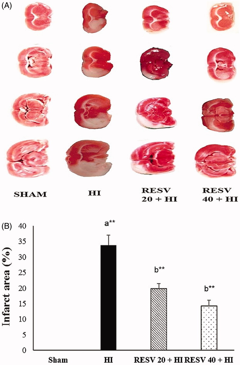
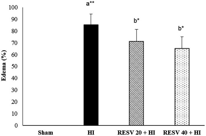
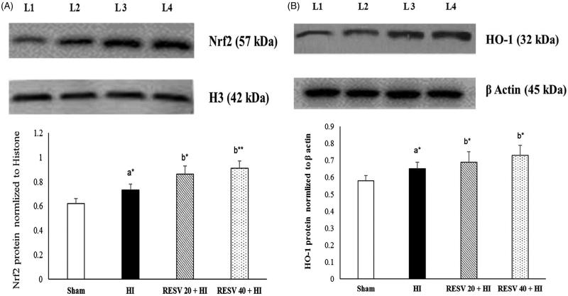
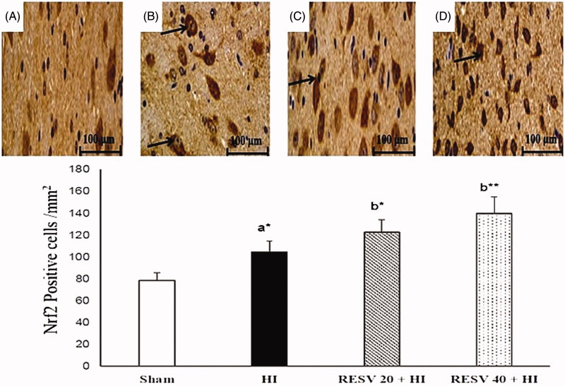
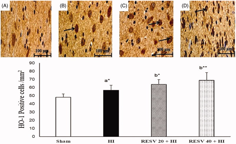
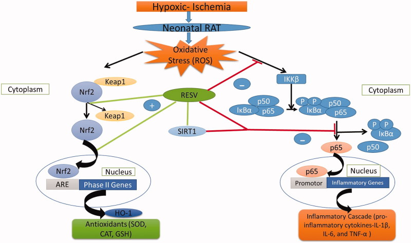
Results: Pre-treatment with RESV (20 or 40) markedly reduced (p < 0.01) the cerebral oedema (86.23-71.26 or 65.24%), infarct area (33.85-19.81 or 14.30%), lipid peroxidation products, inflammatory markers [IL-1β 186-110 or 82; IL-6 255-146 or 103; TNF-α 310-204 or 137; NF-κB 205-115 or 91) p65 subunit] and significantly restored (p < 0.01) the antioxidative status by enhancing the activities of glutathione peroxidase (GPx) 5.22-6.49 or 7.78; catalase (CAT) 51-55 or 59, superoxide dismutase (SOD) 2.5-3.05 or 3.25; through marked upregulation (p < 0.01) of heme oxygenase 1 (HO-1) 0.65-0.69 or 0.73; and nuclear factor erythroid 2 related factor 2 (Nrf2) 0.73-0.86 or 0.91.

Discussion and conclusions: RESV displays its neurotherapeutic potential via upregulating the protein expression of Nrf2 and HO-1 signalling pathway and thereby attenuates oxidative stress and inflammatory response in HI-induced neonatal rats.

Keywords: Lipid peroxidation; infarct area; inflammatory markers; neurotherapeutic; oedema.


Figures

Figure 1.
Figure 1.
Efficacy of RESV on the cerebral infarct area in experimental neonatal rats. Images of coronal transections stained with TTC illustrate numerous white patches which represent the ischemic area (affected area) (HI group; 1B), whereas the normal tissues were red. Data are expressed as the mean ± standard deviation (SD). p value: *p < 0.05, **p < 0.01, Where ‘a’ represent the comparison with the sham control group; ‘b’ represent the comparison with the HI-insulted group.
Figure 2.
Figure 2.
Efficacy of RESV on the cerebral oedema in experimental neonatal rats. Data are expressed as the mean ± standard deviation (SD). Data are expressed as the mean ± standard deviation (SD). p value: *p < 0.05, **p < 0.01, Where ‘a’ represent the comparison with the sham control group; ‘b’ represent the comparison with the HI-insulted group.
Figure 3.
Figure 3.
Efficacy of RESV on the cerebral sections stained with Nissl stain (magnification, ×400) in experimental neonatal rats. Cerebral section of sham control rats (A) shows the normal architecture with many Nissl bodies. Several neuro-morphological changes were noted in hypoxia-ischemia-induced rats (B) with increased pyknotic neurons (indicated by arrows) and less Nissl bodies. Pre-treatment with RESV at 20 (C) and 40 mg/kg (D) for 7 days increased a number of Nissl bodies (viable neurons). Scale bar: 50 µm.
Figure 4.
Figure 4.
Efficacy of RESV on the protein expression of nuclear Nrf2 (A) and cytosolic HO-1 (B) in the cerebral cortex of experimental neonatal rats. Data are expressed as the mean ± standard deviation (SD). p value: *p < 0.05, **p < 0.01, Where ‘a’ represent the comparison with the sham control group; ‘b’ represent the comparison with the HI-insulted group. Lanes 1 (L1): sham control group; Lane 2 (L2): HI group; Lane 3 (L3): RESV 20 + HI group; Lane 4 (L4): RESV 40 + HI group.
Figure 5.
Figure 5.
Efficacy of RESV on the immune-reactivity of Nrf2 (A–D) in the cerebral region of experimental neonatal rats. Relatively less positively stained nuclear Nrf2 was observed in the sham control rats (A). Increased positive staining of cells for nuclear Nrf2 was noted in cerebral tissues (infarct regions) of the HI-induced brain (B). Furthermore, increase in the staining for nuclear Nrf2 was observed (indicated by arrows) in both RESV 20 + HI (C) and 40 + HI groups (D). Scale bar: 100 µm. Data are expressed as the mean ± standard deviation (SD). p value: *p < 0.05, **p < 0.01, Where ‘a’ represent the comparison with the sham control group; ‘b’ represent the comparison with the HI-insulted group.
Figure 6.
Figure 6.
Efficacy of RESV on the immune-reactivity of HO-1 (A–D) in the cerebral region of experimental neonatal rats. In the sham group, only a few cells were positively stained for cytosolic HO-1 (A). In cerebral tissues (infarct regions) of HI-induced rat brains, showed increased positively stained cells for cytosolic HO-1 (B). In the RESV 20 + HI (C) and 40 + HI groups (D), a furthermore increase in the staining for cytosolic HO-1 was observed (indicated by arrows). Scale bar: 100 µm. Data are expressed as the mean ± standard deviation (SD). p value: *p < 0.05, **p < 0.01, Where ‘a’ represent the comparison with the sham control group; ‘b’ represent the comparison with the HI-insulted group.
Figure 7.
Figure 7.
The schematic representation of mechanisms of action of RESV against HIE.

References

    1. Allen KA, Brandon DH. 2011. Hypoxic ischemic encephalopathy: pathophysiology and experimental treatments. Newborn Infant Nurs Rev NAINR. 11:125–133. - PMC - PubMed
    1. Arteaga O, Revuelta M, Montalvo H, Cañavate ML, Alonso-Alconada D, Martinez-Ibargüen A, Hilario E, Alvarez A. 2014. Neuroprotective effect of antioxidants in neonatal rat brain after hypoxia-ischemia: microscopy: advances in scientific research and education. Badajoz, Spain: Formatex Research Center; p. 335–343.
    1. Bastianetto S, Ménard C, Quirion R. 2015. Neuroprotective action of resveratrol. Biochim Biophys Acta. 1852:1195–1201. - PubMed
    1. Block ML, Zecca L, Hong JS. 2007. Microglia-mediated neurotoxicity: uncovering the molecular mechanisms. Nat Rev Neurosci. 8:57–69. - PubMed
    1. Buonocore G, Groenendaal F. 2007. Anti-oxidant strategies. Semin Fetal Neonat Med. 12:287–295. - PubMed

MeSH terms

LinkOut - more resources