Review
. 2019 Mar;105(3):614-624.
doi: 10.1002/cpt.1311. Epub 2019 Jan 11.

Pediatric Anthracycline-Induced Cardiotoxicity: Mechanisms, Pharmacogenomics, and Pluripotent Stem-Cell Modeling

Affiliations
Review

Pediatric Anthracycline-Induced Cardiotoxicity: Mechanisms, Pharmacogenomics, and Pluripotent Stem-Cell Modeling

Anne Tripaydonis et al. Clin Pharmacol Ther. 2019 Mar.

Abstract

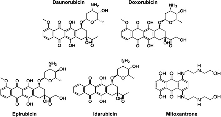
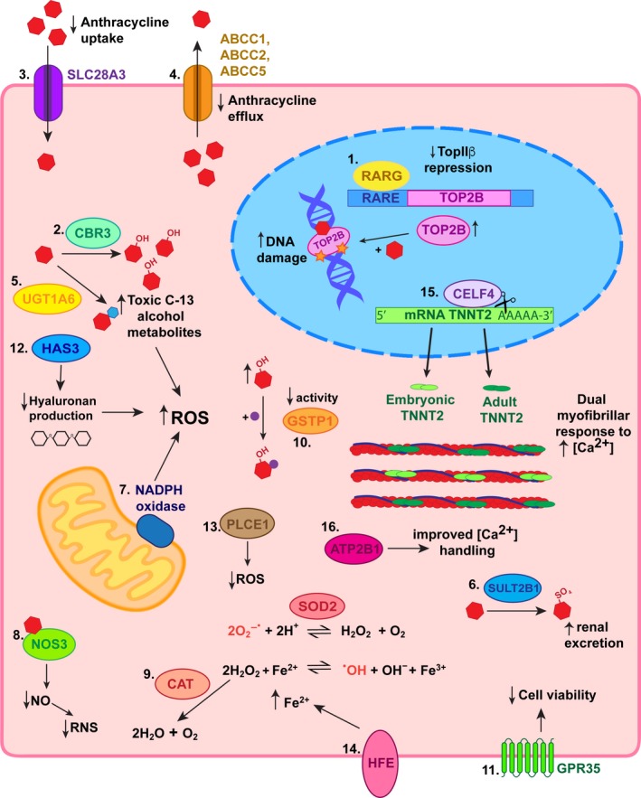
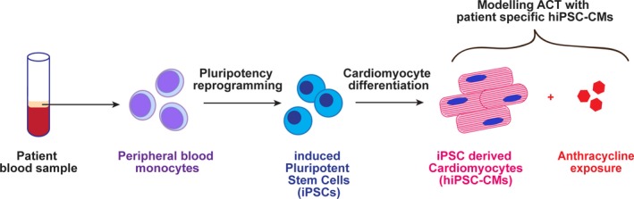
Anthracycline-induced cardiotoxicity (ACT) is a severe adverse drug reaction for a subset of children treated with anthracyclines as part of chemotherapy protocols. The identification of genetic markers associated with increased ACT susceptibility has clinical significance toward improving patient care and our understanding of the molecular mechanisms involved in ACT. Human-induced pluripotent stem cell-derived cardiomyocytes represent a novel approach to determine the pharmacogenomics of ACT and guide the development of genetic screening tests.


Conflict of interest statement

The authors declared no competing interests for this work.

Figures

Figure 1
Figure 1
Chemical structures of clinically approved anthracyclines.
Figure 2
Figure 2
Summary of proposed mechanisms for identified genetic variants associated with anthracycline‐induced cardiotoxicity (ACT) in children. 1. The retinoic acid receptor‐gamma (RARG) variant decreases repression of topoisomerase (TOP)2B such that its levels increase enabling greater amounts of doxorubicin to stabilize its complex with double‐stranded DNA breaks. This leads to increased DNA damage and signals for apoptosis. 2. Carbonyl reductase (CBR)3 variants with increased catalytic activity result in accumulation of toxic C‐13 alcohol metabolites. 3. Decreased anthracycline uptake into cardiomyocytes due to a variant in transporter solute carrier (SLC)28A3 is protective against ACT. 4. The UDP‐glucuronosyltransferase (UGT)1A6 variant increases glucuronidation of anthracyclines resulting in higher levels of toxic metabolites, which predispose patients to ACT. 5. Decreased anthracycline efflux due to polymorphisms in ABC transporters leads to an accumulation of anthracyclines in cardiomyocytes sensitizing them to ACT. 6. Sulfotransferase (SULT)2B1 variant increases sulfonation and therefore renal excretion of anthracyclines and are thus protective against ACT. 7. Mutations in the RAC2 subunit of nicotinamide adenine dinucleotide phosphate (NADPH) oxidase result in increased reactive oxygen species (ROS), which predisposes to ACT. 8. Nitric oxide synthase (NOS)3 variant decreases production of nitric oxide (NO) and consequential reactive nitrogen species (RNS), which culminates in resistance to ACT. 9. A variant resulting in increased cationic amino acid transporter (CAT) expression protects against ACT as hydrogen peroxide is diverted away from conversion to the hydroxyl radical. 10. Conversely, variation causing decreased activity of glutathione s‐transferase p (GSTP)1 results in reduced protection against ROS and therefore increased susceptibility to ACT. 11. A variant in G‐protein‐coupled receptor (GPR)35 has been associated with decreased cell viability upon exposure to anthracyclines. 12. Decreased production of hyaluronan, an antioxidant, due to a polymorphism in hyaluronan synthase (HAS)3 predisposes toward ACT via increased ROS. 13. The phospholipase C epsilon (PLCE)1 variant protects against ACT by reducing the oxidative stress anthracyclines cause. 14. HFE mutations increasing intracellular Fe2+ concentration drive production of hydroxyl radicals. 15. CUGBP Elav‐like family member (CELF)4 variant results in the persistence of alternative splice variants of cardiac troponin T, which are developmentally inappropriate. Co expression of embryonic and adult cardiac troponin T results in a dual capacity of cardiomyocytes to respond to the increasing intracellular calcium levels that occur when anthracyclines are present. 16. An ATP2B1 variant has been associated with ACT resistance potentially as a result of improved calcium handling that optimises sarcomere function during challenge. RARE, retinoic acid receptor element; SOD2, superoxide dismutase II.
Figure 3
Figure 3
Superoxide anion conversion pathway to hydroxyl radical and catalase defense pathway.
Figure 4
Figure 4
Disease modeling anthracycline‐induced cardiotoxicity (ACT) with patient‐specific human‐induced pluripotent stem cells‐derived cardiomyocytes (hiPSCCMs).

References

    1. Miller, K. et al Cancer treatment and survivorship statistics, 2016. CA Cancer J. Clin. 66, 271–289 (2016). - PubMed
    1. Australian Childhood Cancer Registry . A summary of childhood cancer statistics 1983–2014. Queensland: Cancer Council (2014). <https://cancerqld.blob.core.windows.net/site/content/uploads/2017/12/A-s...>. Accessed February 27, 2018.
    1. Smith, L. et al Cardiotoxicity of anthracycline agents for the treatment of cancer: systematic review and meta‐analysis of randomised controlled trials. BMC Cancer 10, 1–14 (2010). - PMC - PubMed
    1. Minotti, G. , Menna, P. , Salvatorelli, E. , Cairo, G. & Gianni, L. Anthracyclines: molecular advances and pharmacologic developments in antitumor activity and cardiotoxicity. Pharmacol. Rev. 56, 185–229 (2004). - PubMed
    1. Jain, K. et al A prospective randomized comparison of epirubicin and doxorubicin in patients with advanced breast cancer. J. Clin. Oncol. 3, 818–826 (1985). - PubMed

MeSH terms