

Washing or filtering of blood products does not improve outcome in a rat model of trauma and multiple transfusion
- PMID: 30461025
- PMCID: PMC7379301
- DOI: 10.1111/trf.15039
Washing or filtering of blood products does not improve outcome in a rat model of trauma and multiple transfusion
Abstract
Background: Transfusion is associated with organ failure and nosocomial infection in trauma patients, which may be mediated by soluble bioactive substances in blood products, including extracellular vesicles (EVs). We hypothesize that removing EVs, by washing or filtering of blood products, reduces organ failure and improves host immune response.
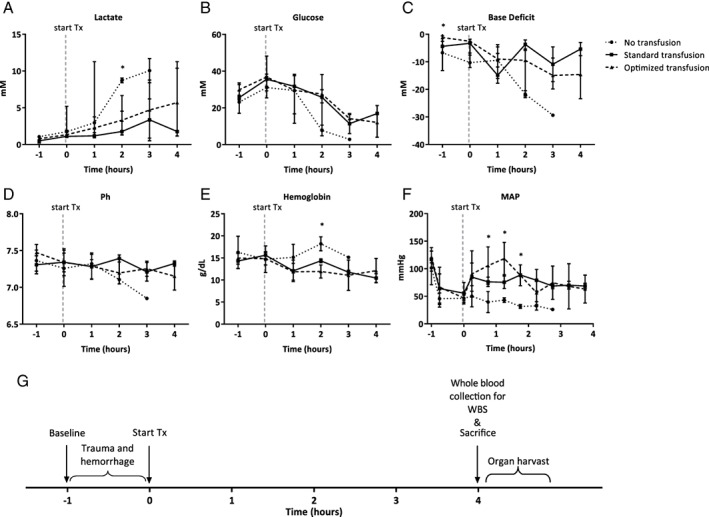
Materials and methods: Blood products were prepared from syngeneic rat blood. EVs were removed from RBCs and platelets by washing. Plasma was filtered through a 0.22-μm filter. Rats were traumatized by crush injury to the intestines and liver, and a femur was fractured. Rats were hemorrhaged until a mean arterial pressure of 40 mm Hg and randomized to receive resuscitation with standard or washed/filtered blood products, in a 1:1:1 ratio. Sham controls were not resuscitated. Ex vivo whole blood stimulation tests were performed and histopathology was done.
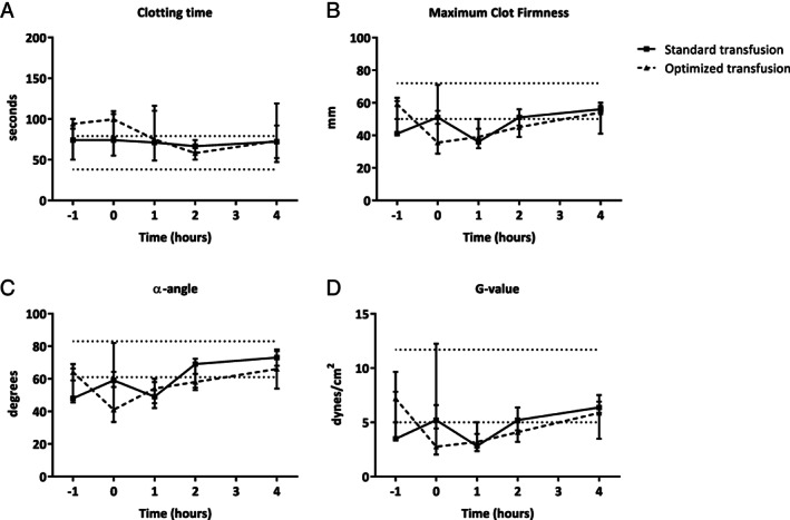
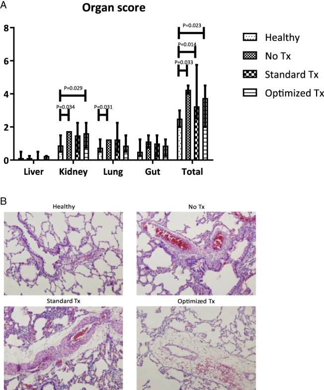
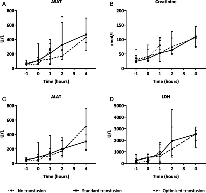
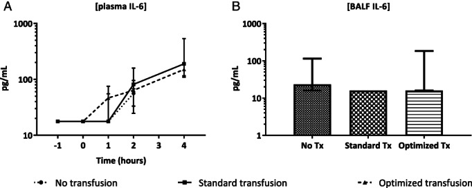
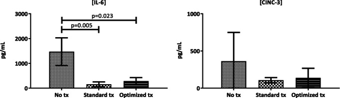
Results: Washing of blood products improved quality metrics compared to standard products. Also, EV levels reduced by 12% to 77%. The coagulation status, as assessed by thromboelastometry, was deranged in both groups and normalized during transfusion, without significant differences. Use of washed/filtered products did not reduce organ failure, as assessed by histopathologic score and biochemical measurements. Immune response ex vivo was decreased following transfusion compared to sham but did not differ between transfusion groups.
Conclusion: Filtering or washing of blood products improved biochemical properties and reduced EV counts, while maintaining coagulation abilities. However, in this trauma and transfusion model, the use of optimized blood components did not attenuate organ injury or immune suppression.
© 2018 The Authors. Transfusion published by Wiley Periodicals, Inc. on behalf of AABB.
Conflict of interest statement
The authors have disclosed no conflicts of interest.
Figures
Similar articles
-
Therapeutic application of recombinant human ADAMTS-13 improves shock reversal and coagulation status in a trauma hemorrhage and transfusion rat model.Intensive Care Med Exp. 2020 Dec 18;8(Suppl 1):42. doi: 10.1186/s40635-020-00328-w. Intensive Care Med Exp. 2020. PMID: 33336308 Free PMC article.
-
Treatment with ddAVP improves platelet-based coagulation in a rat model of traumatic hemorrhagic shock.Trauma Surg Acute Care Open. 2022 Mar 8;7(1):e000852. doi: 10.1136/tsaco-2021-000852. eCollection 2022. Trauma Surg Acute Care Open. 2022. PMID: 35340703 Free PMC article.
-
Implementation of a pediatric trauma massive transfusion protocol: one institution's experience.Transfusion. 2012 Jun;52(6):1228-36. doi: 10.1111/j.1537-2995.2011.03458.x. Epub 2011 Dec 1. Transfusion. 2012. PMID: 22128884
-
Optimal trauma resuscitation with plasma as the primary resuscitative fluid: the surgeon's perspective.Hematology Am Soc Hematol Educ Program. 2013;2013:656-9. doi: 10.1182/asheducation-2013.1.656. Hematology Am Soc Hematol Educ Program. 2013. PMID: 24319247 Review.
-
Resuscitation and transfusion management in trauma patients: emerging concepts.Curr Opin Crit Care. 2012 Dec;18(6):661-70. doi: 10.1097/MCC.0b013e328357b209. Curr Opin Crit Care. 2012. PMID: 22914428 Review.
Cited by
-
Bosutinib reduces endothelial permeability and organ failure in a rat polytrauma transfusion model.Br J Anaesth. 2021 May;126(5):958-966. doi: 10.1016/j.bja.2021.01.032. Epub 2021 Mar 6. Br J Anaesth. 2021. PMID: 33685634 Free PMC article.
-
Shock-Driven Endotheliopathy in Trauma Patients Is Associated with Leucocyte Derived Extracellular Vesicles.Int J Mol Sci. 2022 Dec 15;23(24):15990. doi: 10.3390/ijms232415990. Int J Mol Sci. 2022. PMID: 36555630 Free PMC article.
-
Therapeutic application of recombinant human ADAMTS-13 improves shock reversal and coagulation status in a trauma hemorrhage and transfusion rat model.Intensive Care Med Exp. 2020 Dec 18;8(Suppl 1):42. doi: 10.1186/s40635-020-00328-w. Intensive Care Med Exp. 2020. PMID: 33336308 Free PMC article.
-
Treatment with ddAVP improves platelet-based coagulation in a rat model of traumatic hemorrhagic shock.Trauma Surg Acute Care Open. 2022 Mar 8;7(1):e000852. doi: 10.1136/tsaco-2021-000852. eCollection 2022. Trauma Surg Acute Care Open. 2022. PMID: 35340703 Free PMC article.
-
Use of a high platelet-to-RBC ratio of 2:1 is more effective in correcting trauma-induced coagulopathy than a ratio of 1:1 in a rat multiple trauma transfusion model.Intensive Care Med Exp. 2019 Jul 25;7(Suppl 1):42. doi: 10.1186/s40635-019-0242-5. Intensive Care Med Exp. 2019. PMID: 31346913 Free PMC article.
References
MeSH terms
LinkOut - more resources
Full Text Sources
Medical
