. 2019 Jan;57(1):e23264.
doi: 10.1002/dvg.23264. Epub 2018 Dec 24.

Cadherin-7 mediates proper neural crest cell-placodal neuron interactions during trigeminal ganglion assembly

Affiliations

Cadherin-7 mediates proper neural crest cell-placodal neuron interactions during trigeminal ganglion assembly

Chyong-Yi Wu et al. Genesis. 2019 Jan.

Abstract

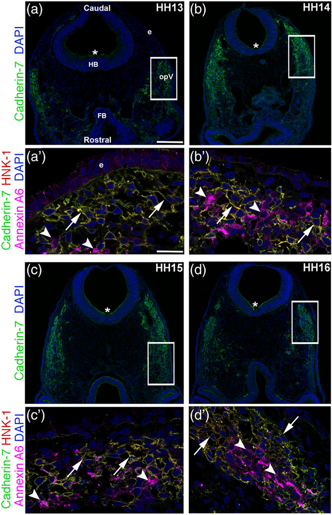
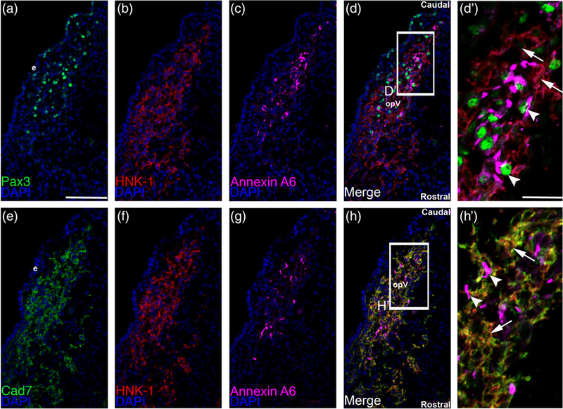
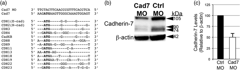
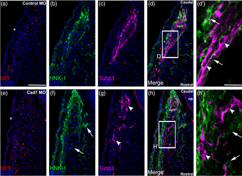
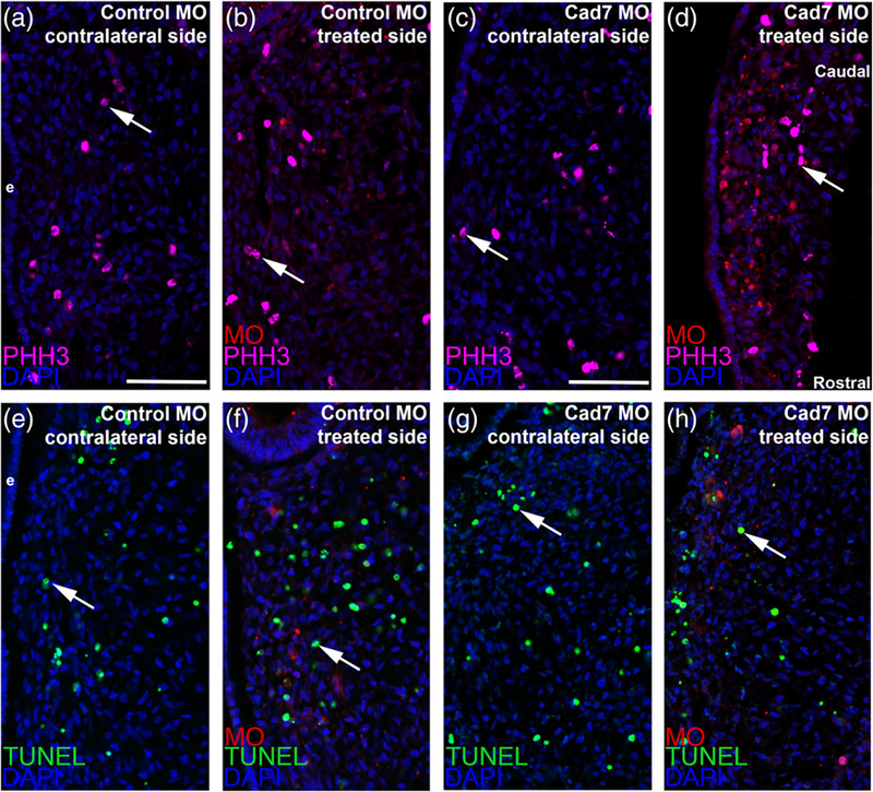
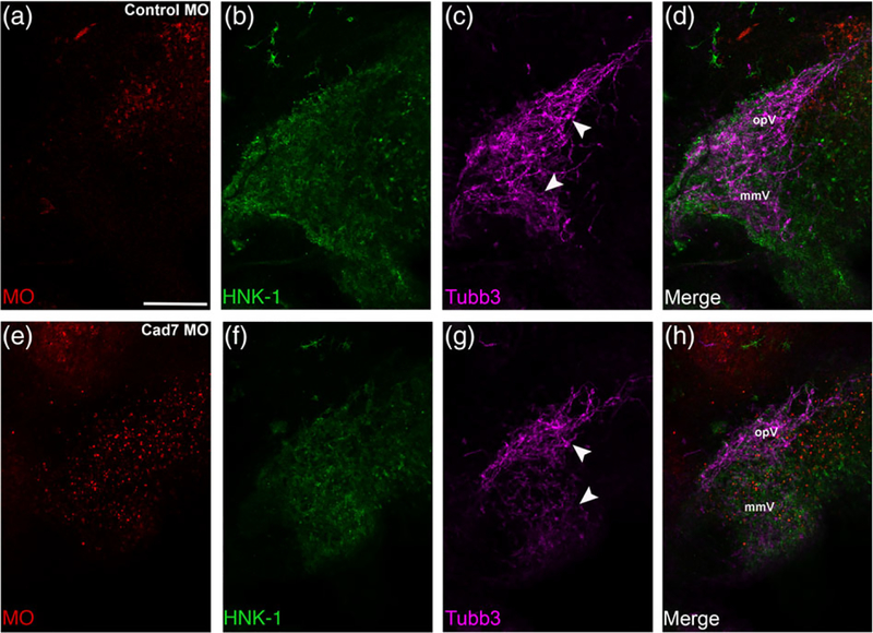
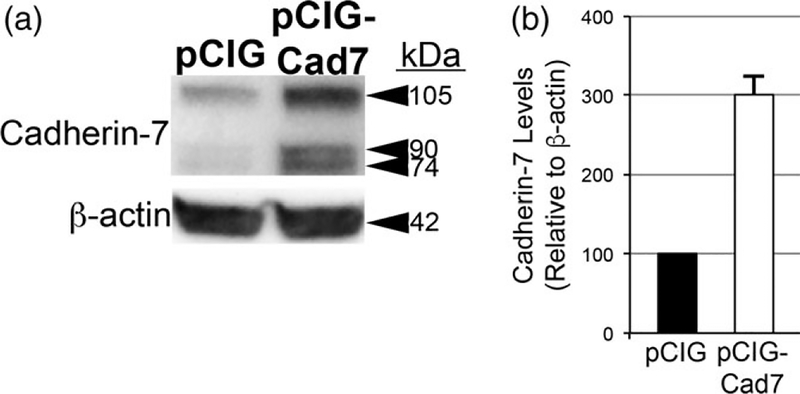
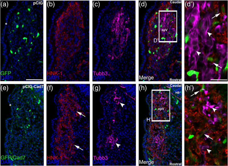
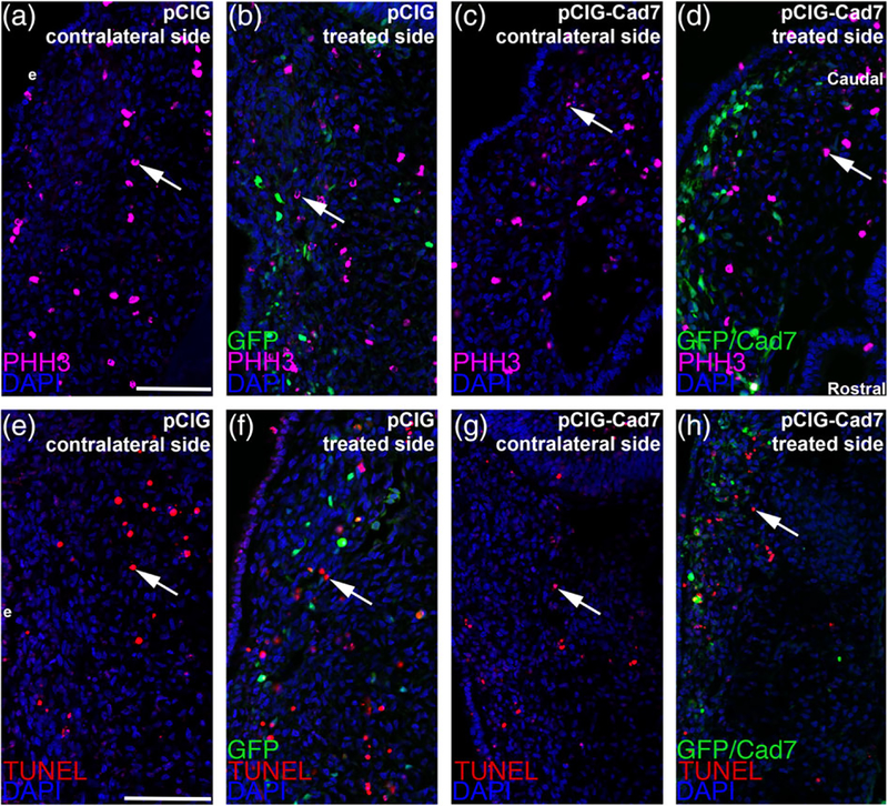
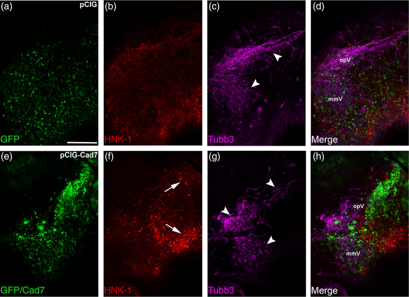
The cranial trigeminal ganglia play a vital role in the peripheral nervous system through their relay of sensory information from the vertebrate head to the brain. These ganglia are generated from the intermixing and coalescence of two distinct cell populations: cranial neural crest cells and placodal neurons. Trigeminal ganglion assembly requires the formation of cadherin-based adherens junctions within the neural crest cell and placodal neuron populations; however, the molecular composition of these adherens junctions is still unknown. Herein, we aimed to define the spatio-temporal expression pattern and function of Cadherin-7 during early chick trigeminal ganglion formation. Our data reveal that Cadherin-7 is expressed exclusively in migratory cranial neural crest cells and is absent from trigeminal neurons. Using molecular perturbation experiments, we demonstrate that modulation of Cadherin-7 in neural crest cells influences trigeminal ganglion assembly, including the organization of neural crest cells and placodal neurons within the ganglionic anlage. Moreover, alterations in Cadherin-7 levels lead to changes in the morphology of trigeminal neurons. Taken together, these findings provide additional insight into the role of cadherin-based adhesion in trigeminal ganglion formation, and, more broadly, the molecular mechanisms that orchestrate the cellular interactions essential for cranial gangliogenesis.

Keywords: Cadherin-7; neural crest; placodal neurons; trigeminal ganglion.


Figures

FIGURE 1
FIGURE 1
Cadherin-7 protein is observed in migratory neural crest cells contributing to the trigeminal ganglion. Representative transverse sections taken at the axial level of the forming ophthalmic lobe of the trigeminal ganglion over four chick embryo stages (HH13–16) followed by immunohistochemistry for Cadherin-7 (green), HNK-1 (red, labels neural crest cells), and Annexin A6 (purple, labels placodal neurons). (a–d) Lower magnification images show the entire transverse section and reveal Cadherin-7 immunoreactivity, with rostral and caudal orientation, the location of the forebrain (FB), hindbrain (HB), and ectoderm (e), and the ophthalmic lobe of the trigeminal (opV), indicated in (a) and applicable to (b–d). Asterisk (*) in (a–d) reveals Cadherin-7 immunoreactivity in the neural tube. Higher magnification images (a′–d′) of the boxed area in (a–d) show Cadherin-7-and HNK-1-double-positive neural crest cells at all stages (arrows), whereas Annexin A6-positive placodal neurons are devoid of Cadherin-7 (arrowheads). DAPI (blue) labels cell nuclei. Scale bar in (a) is 100 μm and applicable to (b–d), while scale bar in (a′) is 5 μm and applicable to (b′–d′)
FIGURE 2
FIGURE 2
Cadherin-7 protein localizes to HNK1-positive neural crest cells but not Pax3-positive placode cells or placodal neurons. Representative serial/adjacent transverse sections taken at the axial level of the forming ophthalmic lobe of the trigeminal ganglion at HH15 followed by triple-label immunohistochemistry for either Pax3 (a, green), HNK-1 (b, red), and Annexin A6 (c, purple), or Cadherin-7 (e, green), HNK-1 (f, red), and Annexin A6 (g, purple). (d′) Higher magnification image of the boxed region in (d) shows placode cells or placodal neurons (Pax-3- and Annexin A6-positive cells, arrowheads) that are not labeled with HNK-1 and thus are different from HNK-positive neural crest cells (arrows). (h′) Higher magnification image of the boxed region in (h) reveals Cadherin-7 immunoreactivity solely in HNK-1-positive neural crest cells (arrows), while Annexin A6-positive placodal neurons (arrowheads) are devoid of Cadherin-7. Rostral and caudal orientation and the ophthalmic lobe of the trigeminal (opV) are indicated in (d) and (h). DAPI (blue) labels cell nuclei. e = ectoderm. Scale bar in (a) is 67 μm and applicable to (b–h), while scale bar in (d′) is 20 μm and applicable to (h′)
FIGURE 3
FIGURE 3
Knockdown of Cadherin-7 with a translation-blocking morpholino antisense oligonucleotide targeting Cadherin-7 effectively reduces Cadherin-7 protein in migratory neural crest cells contributing to the trigeminal ganglion. (a) Comparison of Cadherin-7 MO (Cad7 MO) sequence to the targeting sequence in the Cadherin-7 transcript and to other Cadherin transcripts with expression patterns in premigratory and/or migratory neural crest cells, the neural tube, and in the region of the forming trigeminal ganglion. (b) Premigratory neural crest cells were electroporated at the 2–3ss with either the Cadherin-7 morpholino (Cad7 MO) to allow for depletion of Cadherin-7 protein in migratory neural crest cells, or a 5 bp mismatch Cadherin-7 control MO (Ctrl MO). Embryos were re-incubated to HH15–17 after which time the trigeminal ganglion-forming region on the electroporated side of the embryo was dissected out of the embryo and pooled for lysate preparation. Immunoblotting for Cadherin-7 and β-actin (control) was performed as in (Shah, Schiffmacher, & Taneyhill, 2017), with a representative immunoblot shown. (c) Knockdown efficiency of the Cad7 MO was assessed as previously described (Shah et al., 2017), with graph revealing results of immunoblot analysis as determined by normalizing Cadherin-7 to β-actin and calculating the reduction in this normalized ratio from that obtained for the control MO-treated lysate (arbitrarily set to 1, n = 2). The mean and standard error of the mean are shown. A 50% knockdown in Cadherin-7 protein levels is noted in the Cadherin-7 MO-treated lysate compared to the control MO-treated lysate
FIGURE 4
FIGURE 4
Morpholino-mediated depletion of Cadherin-7 from migratory cranial neural crest cells alters the distribution of neural crest cells and placodal neurons within the forming trigeminal ganglion. Representative transverse sections taken at the axial level of the forming ophthalmic lobe of the trigeminal ganglion after electroporation of a 5 bp mismatch control Cadherin-7 morpholino (control MO, a–d′) or Cadherin-7 MO (Cad7 MO, e–h′) into premigratory neural crest cells at the 3ss followed by immunohistochemistry for HNK-1 (green) and Tubb3 (purple) at HH15. (d′, h′) Higher magnification images of the boxed regions in (d, h). (a–d′) Control MO (a)-containing neural crest cells (b) coalesce with placodal neurons (c, d). At higher magnification (d′), HNK-1-positive neural crest cells (arrows) surround Tubb3-positive placodal neurons (arrowheads), many of which are already forming neurites or adopting the bipolar morphology associated with neuronal maturation. Conversely, a trigeminal ganglion containing the Cadherin-7 MO (e) in the neural crest (f) reveals differences in neural crest cell (f, h, arrows) and placodal neurons (g, h, arrowheads) distribution within the anlage, with neural crest cells sometimes appearing more dispersed compared to control. At higher magnification (h′), it is apparent that neural crest cells still surround the placodal neurons (arrows), but the shape adopted by the placodal neurons is aberrant, with neurons appearing round (arrowheads). Rostral and caudal orientation, the hindbrain (HB), and the ophthalmic lobe of the trigeminal ganglion (opV) are indicated in (d) and (h). DAPI (blue) labels cell nuclei. e = ectoderm. Scale bar in (a) is 67 μm and applicable to (b–h), while scale bar in (d′) is 20 μm and applicable to (h′)
FIGURE 5
FIGURE 5
Electroporation of either the control or Cadherin-7 morpholino does not alter cell proliferation or cell death in the trigeminal ganglionic anlage. Representative transverse sections taken at the axial level of the forming ophthalmic lobe of the trigeminal ganglion after electroporation of a 5 bp mismatch control Cadherin-7 morpholino (control MO: a, b, e, f) or Cadherin-7 morpholino (Cad7 MO: c, d, g, h) into premigratory neural crest cells at the 3ss followed by immunohistochemistry for phospho-histone H3 (PHH3, a–d) or TUNEL (e–h) at HH15. Contralateral (a, c, e, g) and morpholino-treated (b, d, f, h) sides are shown to provide a means of comparison. Arrows indicate PHH3 (a–d)- and TUNEL (e–h)-positive nuclei, with a comparable number noted in the presence of either morpholino relative to the contralateral control side of the electroporated embryo. DAPI (blue) labels cell nuclei. Ectoderm (e) is oriented to the left within each image panel and may not be visible in the field of view for some images. Rostral and caudal orientation is shown in (d) and applies to all images. Scale bar in (a) is 67 μm and applies to (b, e, f), while scale bar in (c) is 50 μm and applies to (d, g, h)
FIGURE 6
FIGURE 6
Cadherin-7 depletion in migratory neural crest cells alters the gross morphology of the trigeminal ganglion. Representative lateral views (optical section) of the forming trigeminal ganglion in an HH15 chick head after electroporation of a 5 bp mismatch control Cadherin-7 MO (control MO, a–d) or Cadherin-7 MO (Cad7 MO, e–h) at the 3ss, followed by whole-mount immunohistochemistry for HNK-1 (green) and Tubb3 (purple). Merge images are shown in (d, h). A trigeminal ganglion electroporated with the control MO in neural crest cells (a) exhibits a bilobed morphology, with neural crest cells (b) condensing with placodal neurons (c, arrowheads). A trigeminal ganglion electroporated with the Cadherin-7 MO in the neural crest (e) possesses neural crest cells that migrate to the anlage (f) but placodal neurons do not condense properly (g, arrowheads), leading to an aberrant ganglion shape relative to control. The ophthalmic (opV) and maxillomandibular (mmV) lobes of the trigeminal ganglion are indicated in (d) and (h). Scale bar in (a) is 200 μm and applies to all images
FIGURE 7
FIGURE 7
Overexpression of Cadherin-7 effectively increases Cadherin-7 protein in neural crest cells contributing to the trigeminal ganglion. (a) Premigratory neural crest cells were electroporated at 2–3ss with either a Cadherin-7 expression construct (pCIG-Cad7) to allow for overexpression of Cadherin-7 protein in migratory neural crest cells, or the control vector (pCIG). Embryos were re-incubated HH15–17 after which time the trigeminal ganglion-forming region on the electroporated side of the embryo was dissected out of the embryo and pooled for lysate preparation. Immunoblotting for Cadherin-7 and β-actin (control) was performed as in (Shah et al.,2017), with a representative immunoblot shown. (b) Overexpression efficiency was assessed as previously described (Shah et al., 2017), with graph indicating results of immunoblot analysis as determined by normalizing Cadherin-7 to β-actin and calculating the increase in this normalized ratio from that obtained for the pCIG-treated lysate (arbitrarily set to 1, n = 2). The mean and standard error of the mean are shown. A 200% increase in Cadherin-7 protein levels is noted in the pCIG-Cad7-treated lysate compared to the control pCIG-treated lysate
FIGURE 8
FIGURE 8
Overexpression of Cadherin-7 in migratory cranial neural crest cells alters the distribution of neural crest cells and placodal neurons within the forming trigeminal ganglion. Representative transverse sections taken at the axial level of the forming ophthalmic lobe of the trigeminal ganglion after electroporation of the pCIG control vector (pCIG, a–d′) or the pCIG-Cadherin-7 vector (pCIG-Cad7, e–h′) into premigratory neural crest cells at the 3ss followed by immunohistochemistry for HNK-1 (red) and Tubb3 (purple) at HH15. The pCIG vector contains an IRES-GFP cassette to label electroporated cells. (d′, h′) Higher magnification images of the boxed regions in (d, h). (a–d′) pCIG control vector (a)-containing neural crest cells (b) coalesce with placodal neurons (c, d). At higher magnification (d′), HNK-1-positive neural crest cells (arrows) form corridors around Tubb3-positive placodal neurons (arrowheads), many of which are elaborating neurites indicative of neuronal maturation. On the other hand, neural crest cells with elevated levels of Cadherin-7 protein (e) migrate to the ganglionic anlage (f, arrows) but likely localize there incorrectly given the inappropriate positioning of placodal neurons (g, arrowheads). At higher magnification (h′), neural crest cells are noted around the placodal neurons (arrows), but placodal neuron morphology is abnormal, with neurons appearing round and/or misshapen (arrowheads). Rostral and caudal orientation, the hindbrain (HB), and the ophthalmic lobe of the trigeminal (opV) are indicated in (d) and (h). DAPI (blue) labels cell nuclei. e = ectoderm. Scale bar in (a) is 50 μm and applicable to (b–h), while scale bar in (d′) is 20 μm and applicable to (h′)
FIGURE 9
FIGURE 9
Electroporation of expression constructs does not alter cell death or cell proliferation in the trigeminal ganglionic anlage. Representative transverse sections taken at the axial level of the forming ophthalmic lobe of the trigeminal ganglion after electroporation of the pCIG control vector (pCIG: a, b, e, f) or pCIG-Cadherin-7 vector (pCIG-Cad7: c, d, g, h) into premigratory neural crest cells at the 3ss followed by immunohistochemistry for phospho-histone H3 (PHH3, a–d) or TUNEL (e–h) at HH15. Contralateral (a, c, e, g) and expression vector-treated (b, d, f, h) sides are shown to provide a means of comparison. Arrows indicate PHH3 (a–d)-and TUNEL (e–h)-positive nuclei, with a comparable number noted in the presence of either expression construct relative to the contralateral control side of the electroporated embryo. DAPI (blue) labels cell nuclei. Ectoderm (e) is oriented to the left within each image panel. Rostral and caudal orientation is shown in (d) and applies to all images. Scale bar in (a) is 60 μm and applies to (b–d), while scale bar in (e) is 60 μm and is applies to (f–h)
FIGURE 10
FIGURE 10
Elevated levels of Cadherin-7 in migratory neural crest cells alter the gross morphology of the trigeminal ganglion. Representative lateral views (optical section) of the forming trigeminal ganglion in an HH15 chick head after electroporation of the pCIG control vector (pCIG, a–d) or the pCIG-Cadherin-7 vector (pCIG-Cad7, e–h) at the 3ss, followed by whole-mount immunohistochemistry for HNK-1 (red) and Tubb3 (purple). Merge images are shown in (d, h). A trigeminal ganglion electroporated with the control pCIG vector in neural crest cells (a) exhibits a bilobed morphology, with neural crest cells (b) coalescing with placodal neurons (c, arrowheads). A trigeminal ganglion electroporated with pCIG-Cad7 in the neural crest (e) possesses neural crest cells that migrate to the anlage, but these cells appear to aggregate together, thus altering their general distribution in the anlage (e, f, arrows). Placodal neurons are also affected, exhibiting a less compact appearance (g, arrowheads). Together, this leads to an abnormal ganglion shape relative to control. The ophthalmic (opV) and maxillomandibular (mmV) lobes of the trigeminal ganglion are indicated in (d) and (h). Scale bar in (a) is 200 μm and applies to all images

Similar articles

Cited by

References

    1. Baker CV, & Bronner-Fraser M (2001). Vertebrate cranial placodes I. Embryonic induction. Developmental Biology, 232, 1–61. - PubMed
    1. Basu R, Duan X, Taylor MR, Martin EA, Muralidhar S, Wang Y, … Williams ME (2017). Heterophilic type II Cadherins are required for high-magnitude synaptic potentiation in the hippocampus. Neuron, 96, 160–176e8. - PMC - PubMed
    1. Breau MA, & Schneider-Maunoury S (2015). Cranial placodes: Models for exploring the multi-facets of cell adhesion in epithelial rearrangement, collective migration and neuronal movements. Developmental Biology, 401, 25–36. - PubMed
    1. Bronner ME, & Simoes-Costa M (2016). The neural crest migrating into the twenty-first century. Current Topics in Developmental Biology, 116, 115–134. - PMC - PubMed
    1. Bronner-Fraser M (1986). Analysis of the early stages of trunk neural crest migration in avian embryos using monoclonal antibody HNK-1. Developmental Biology, 115, 44–55. - PubMed

Publication types

LinkOut - more resources