. 2019 Feb 1;316(2):L321-L333.
doi: 10.1152/ajplung.00056.2018. Epub 2018 Nov 21.

BPIFA1 regulates lung neutrophil recruitment and interferon signaling during acute inflammation

Affiliations

BPIFA1 regulates lung neutrophil recruitment and interferon signaling during acute inflammation

Clemente J Britto et al. Am J Physiol Lung Cell Mol Physiol. .

Abstract

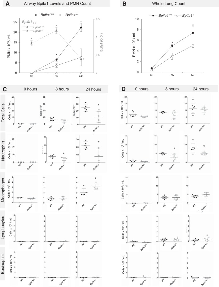
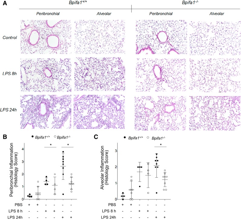
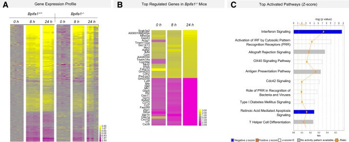
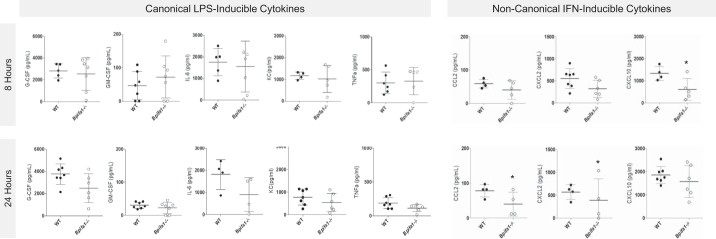
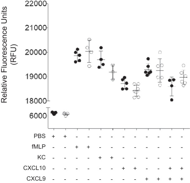
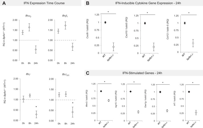
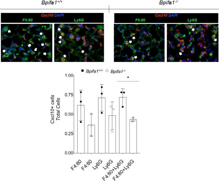
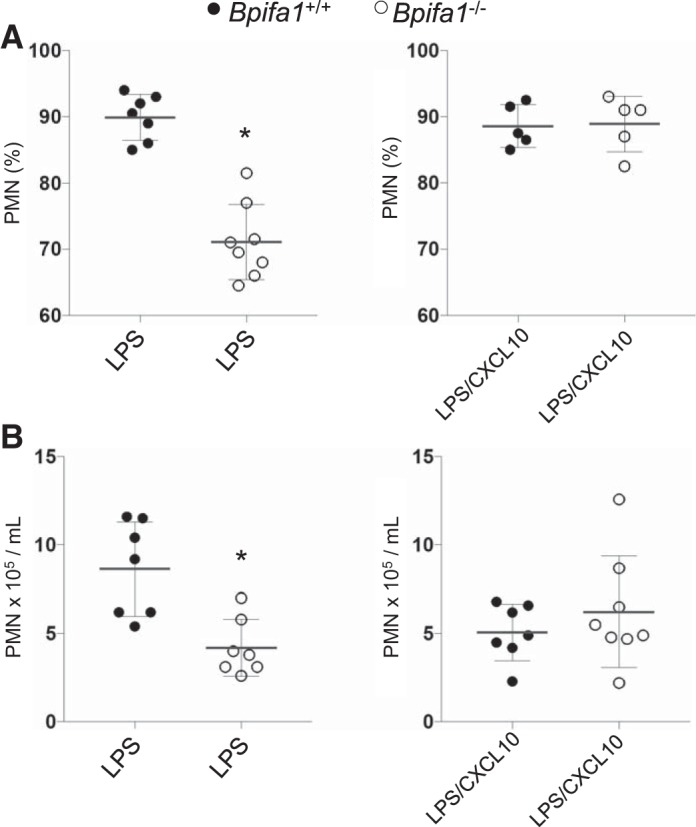
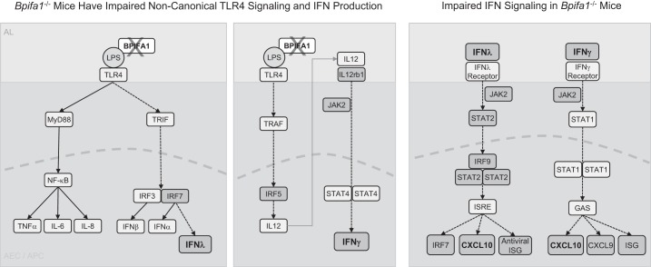
Bpifa1 (BPI fold-containing group A member 1) is an airway host-protective protein with immunomodulatory properties that binds to LPS and is regulated by infectious and inflammatory signals. Differential expression of Bpifa1 has been widely reported in lung disease, yet the biological significance of this observation is unclear. We sought to understand the role of Bpifa1 fluctuations in modulating lung inflammation. We treated wild-type (WT) and Bpifa1-/- mice with intranasal LPS and performed immunological and transcriptomic analyses of lung tissue to determine the immune effects of Bpifa1 deficiency. We show that neutrophil (polymorphonuclear cells, PMNs) lung recruitment and transmigration to the airways in response to LPS is impaired in Bpifa1-/- mice. Transcriptomic analysis revealed a signature of 379 genes that differentiated Bpifa1-/- from WT mice. During acute lung inflammation, the most downregulated genes in Bpifa1-/- mice were Cxcl9 and Cxcl10. Bpifa1-/- mice had lower bronchoalveolar lavage concentrations of C-X-C motif chemokine ligand 10 (Cxcl10) and Cxcl9, interferon-inducible PMN chemokines. This was consistent with lower expression of IFNγ, IFNλ, downstream IFN-stimulated genes, and IFN-regulatory factors, which are important for the innate immune response. Administration of Cxcl10 before LPS treatment restored the inflammatory response in Bpifa1-/- mice. Our results identify a novel role for Bpifa1 in the regulation of Cxcl10-mediated PMN recruitment to the lungs via IFNγ and -λ signaling during acute inflammation.


Conflict of interest statement

No conflicts of interest, financial or otherwise, are declared by the authors.

Figures

Fig. 1.
Fig. 1.
BPI fold-containing group A member 1-deficient (Bpifa1−/−) mice have impaired lung neutrophilic inflammation after lipopolysaccharide (LPS). A: neutrophils (black lines) in bronchoalvolar lavage (BAL) fluid from Bpifa1+/+ [wild-type (WT), ●], Bpifa1−/− mice (○), and Bpifa1 protein in BAL (▲) after intranasal LPS (5 μg/mouse, from Pseudomonas aeruginosa). B: neutrophils in lung digests after intranasal LPS. C: total BAL cell counts in WT (●) and Bpifa1−/− mice (○). D: total lung cell counts in WT and Bpifa1−/− mice. O.D., optical density; PMN, polymorphonuclear neutrophils. Mann-Whitney test: *P < 0.05. Horizontal lines indicate mean values. Data represent 3 experiments; n = 4–7 mice/group.
Fig. 2.
Fig. 2.
BPI fold-containing group A member 1-deficient (Bpifa1−/−) mice have limited peribronchial and alveolar inflammation in response to lipopolysaccharide (LPS). A: tissue sections of paraffin-embedded lung tissue stained with hematoxylin-eosin from wild-type (WT; left) and Bpifa1−/− mice (right) after treatment with LPS (LPS 8 h, LPS 24 h) or PBS (Control). Images obtained with optical microscope at ×20 magnification. B: assessment of peribronchial inflammation in WT (●) and Bpifa1−/− mice (○) based on histology score. C: assessment of alveolar inflammation in WT and Bpifa1−/− mice based on histology score. Each data point represents the mean inflammation score of 10 random fields scored within the same section. Black horizontal lines indicate mean values. Generalized linear mixed model: *P < 0.05. Data represent 2 experiments; n = 4–7 mice/group.
Fig. 3.
Fig. 3.
Gene expression profile of BPI fold-containing group A member 1-deficient (Bpifa1−/−) and wild-type (WT) mice during acute lung inflammation. A: whole lung microarray heatmap showing differential gene expression in WT (Bpifa1+/+) and Bpifa1−/− mice after intranasal instillation of lipopolysaccharide (LPS) [2-way ANOVA, multiple hypothesis testing corrected using Benjamini-Hochberg false discovery rate (FDR) set at <0.05]. Adjacent color scale in log base-2 scale. Yellow denotes increase over the geometric mean of samples; purple denotes decrease. B: microarray heatmap showing the top 20 most up/downregulated genes in Bpifa1−/− mice after intranasal instillation of LPS (2-way ANOVA, multiple-hypothesis testing corrected using Benjamini-Hochberg FDR set at <0.05). Adjacent color scale in log base-2 scale: yellow denotes increase over the geometric mean of samples; purple denotes decrease relative to WT expression. C: activated canonical pathways based on gene expression profile. IRF, interferon regulatory factors. Z-scores (bars) represent the strength of the association between the regulated genes within the canonical pathway. Negative Z-score represents statistically significant downregulation of the genes within the pathway relative to WT expression (P < 0.05); positive Z-score represents upregulation (P < 0.05). Ratio (yellow line) represents the fraction of the genes associated with the pathway that are significantly regulated in Bpifa1−/− mice; n = 5–6 mice/group.
Fig. 4.
Fig. 4.
C-X-C motif chemokine ligand 10 (CXCL10) is decreased in BPI fold-containing group A member 1-deficient (Bpifa1−/−) airways during early lung inflammation. Bronchoalveolar lavage from Bpifa1+/+ [wild-type (WT), ●] and Bpifa1−/− mice (○) was collected 8 h (top row) and 24 h (bottom row) after intranasal lipopolysaccharide (LPS). Cytokine concentrations measured by multiplexed Luminex assay. Additional cytokines tested without significant difference: interferon (IFN)γ, interleukin (IL)-1α, IL-1β, IL-2, IL-4, IL-5, IL-7, IL-9, IL-10, IL-15, IL-17, C-C motif chemokine ligand 5 (CCL5), macrophage inflammatory protein (MIP)-1α, MIP-1β. Leukotriene B4 (LTB4) was not significantly different. TNFα, tumor necrosis factor-α; G-CSF, granulocyte colony-stimulating factor; GM-CSF, granulocyte-macrophage colony-stimulating factor; KC, keratinocyte chemoattractant; n = 4–7 mice/group. Mann-Whitney test: *P < 0.05. Horizontal lines indicate mean value. Data represent 2 experiments.
Fig. 5.
Fig. 5.
BPI fold-containing group A member 1-deficient (Bpifa1−/−) polymorphonuclear neutrophils (PMN) do not have impaired chemotaxis response. PMN from wild-type (WT) and Bpifa1−/− mice were isolated from bone marrrow. Chemotaxis assays were performed using a modified Boyden technique. WT and Bpifa1−/− PMN were stimulated with PMN chemoattractant N-formyl-methionyl-leucyl-phenylalanine (fMLP; 105 M), keratinocyte chemoattractant (KC; 50 ng/ml), C-X-C motif chemokine ligand 10 (Cxcl10; 100 ng/ml), Cxcl9 (50 ng/ml), and combination of Cxcl10 and Cxcl9. Total PMN migration represented as relative fluorescence units (RFU). ●, WT PMN; ○, Bpifa1−/− PMN; n = 4–6 wells/group. Generalized linear mixed model: no significant differences observed. Horizontal bars indicate mean values. Data represent 2 experiments.
Fig. 6.
Fig. 6.
Interferon (IFN) and IFN-stimulated gene (ISG) expression in BPI fold-containing group A member 1-deficient (Bpifa1−/−) lungs during acute inflammation. RNA was extracted from whole lung lysates from wild-type (WT) and Bpifa1−/− naive mice (0 h) and treated mice 8 and 24 h after lipopolysaccharide (LPS; 8 h, 24 h). Differences in gene expression were assessed by RT-PCR. A: time course of IFN mRNA expression in Bpifa1−/− mice. Relative expression (RQ) in Bpifa1−/− mice (◇) vs. WT gene expression (WT expression = 1, marked with horizontal dashed line). RQ values lower than 1 denote lower gene expression relative to WT. B: IFN-inducible cytokine gene expression at 24 h: relative expression of cytokine gene mRNA in Bpifa1−/− lungs (◇) relative to WT (◆). Cxcl, C-X-C motif chemokine ligand; Ccl, C-C motif chemokine ligand; tubb5, tubulin-β5. C: IFN-stimulated gene (ISG) expression at 24 h: relative expression of ISG mRNA in Bpifa1−/− lungs (◇) relative to WT (◆). Data represent 2 experiments. Each data point represents the mean mRNA expression of Bpifa1−/− mice within each group. Generalized linear mixed model: n = 4–6 mice/group, *P < 0.05.
Fig. 7.
Fig. 7.
C-X-C motif chemokine ligand 10 (Cxcl10) induction in myeloid cells during inflammation is impaired in BPI fold-containing group A member 1-deficient (Bpifa1−/−) mice. Top: lung sections from wild-type (WT) and Bpifa1−/− mice 24 h after intranasal instillation of lipopolysaccharide (LPS). Tissue sections were stained with antibodies against Cxcl10 (red) and F4:80, a marker for macrophages, or Ly6G, a marker that primarily stains polymorphonuclear neutrophils (PMN; both in green). Cell nuclei were stained with DAPI (blue); colocalization of Cxcl10 and cell markers is shown in yellow (white arowheads). Bottom: ratios of Cxcl10+ stained cells to total Ly6G+, F4:80+, or combined Ly6G+ plus F4:80+ cells in tissue sections from mice in these experiments. Each data point represents the average number of cells per field in 10 random fields at ×20 magnification. ●, WT values; ○, Bpifa1−/− values; n = 3 mice/group. Generalized linear mixed model: *P < 0.05. Data represent 2 experiments.
Fig. 8.
Fig. 8.
Reconstitution of C-X-C motif chemokine ligand 10 (Cxcl10) in BPI fold-containing group A member 1-deficient (Bpifa1−/−) mice restores lung polymorphonuclear neutrophils (PMN). Wild-type (WT) and Bpifa1−/− mice received intraperitoneal Cxcl10 before treatment with lipopolysaccharide (LPS). PMN bronchoalveolar lavage (BAL) counts were assessed at 24 h. A: %PMN isolated in BAL fluid (BALF). B: absolute PMN count in BALF after LPS. Bpifa1+/+, ●; Bpifa1−/−, ○; LPS, 5 μg/mouse, from Pseudomonas aeruginosa PAO1; Cxcl10, 0.6 μg/mouse; n = 5–8 mice/group. Mann-Whitney test: *P < 0.05. Horizontal lines indicate mean values. Data represent 2 experiments.
Fig. 9.
Fig. 9.
Diagram: BPI fold-containing group A member 1 (Bpifa1) regulation of noncanonical Toll-like receptor 4 (TLR4) and interferon (IFN) signaling. Left: Bpifa1 is a lipopolysaccharide (LPS)-binding molecule and is likely to influence inflammatory signaling by modulating LPS-TLR4 interactions. In the absence of Bpifa1, impaired noncanonical TLR4 signaling through TIR domain-containing adapter-inducing INFβ1 (TRIF) results in decreased interferon regulatory factor 7 (IRF7) expression, leading to decreased IFNλ expression. Canonical LPS-TLR4 activation and cytokine expression is not affected by Bpifa1 deficiency. Middle: Bpifa1 binding to LPS facilitates immune signaling through TNF receptor associated factors (TRAF), which in turn activates IRF5 to induce interleukin (IL)-12 expression, inducing IFNγ. During Bpifa1 deficiency, key components of this pathway are downregulated (IL-12rb1, Jak2, Irf5), resulting in decreased IFNγ expression. Right: decreased expression of IFNγ and IFNλ during Bpifa1 deficiency causes downstream downregulation of Irf9, Irf7, Cxcl10, Cxcl9 and IFN-stimulated genes (ISG). Shaded boxes denote lower gene expression in our model. Dashed arrows represent impaired signaling pathways in our model. AEC, airway epithelial cell; APC, antigen-presenting cell; AL, airway lumen; NF-κB, nuclear factor-κB; TNFα, tumor necrosis factor-α; JAK, Janus kinase; STAT, signal transducer and activator of transcription; CXCL, C-X-C motif chemokine ligand.

Similar articles

Cited by

References

    1. Aichele D, Schnare M, Saake M, Röllinghoff M, Gessner A. Expression and antimicrobial function of bactericidal permeability-increasing protein in cystic fibrosis patients. Infect Immun 74: 4708–4714, 2006. doi:10.1128/IAI.02066-05. - DOI - PMC - PubMed
    1. Akram KM, Moyo NA, Leeming GH, Bingle L, Jasim S, Hussain S, Schorlemmer A, Kipar A, Digard P, Tripp RA, Shohet RV, Bingle CD, Stewart JP. An innate defense peptide BPIFA1/SPLUNC1 restricts influenza A virus infection. Mucosal Immunol 11: 71–81, 2018. doi:10.1038/mi.2017.45. - DOI - PubMed
    1. Allard JB, Poynter ME, Marr KA, Cohn L, Rincon M, Whittaker LA. Aspergillus fumigatus generates an enhanced Th2-biased immune response in mice with defective cystic fibrosis transmembrane conductance regulator. J Immunol 177: 5186–5194, 2006. doi:10.4049/jimmunol.177.8.5186. - DOI - PubMed
    1. Barnes FA, Bingle L, Bingle CD. Pulmonary genomics, proteomics, and PLUNCs. Am J Respir Cell Mol Biol 38: 377–379, 2008. doi:10.1165/rcmb.2007-0388TR. - DOI - PubMed
    1. Bauer Y, Tedrow J, de Bernard S, Birker-Robaczewska M, Gibson KF, Guardela BJ, Hess P, Klenk A, Lindell KO, Poirey S, Renault B, Rey M, Weber E, Nayler O, Kaminski N. A novel genomic signature with translational significance for human idiopathic pulmonary fibrosis. Am J Respir Cell Mol Biol 52: 217–231, 2015. doi:10.1165/rcmb.2013-0310OC. - DOI - PMC - PubMed

Publication types

LinkOut - more resources