. 2019 Jan 1;121(1):140-151.
doi: 10.1152/jn.00004.2018. Epub 2018 Nov 21.

Interaction between TRPV1-expressing neurons in the hypothalamus

Affiliations

Interaction between TRPV1-expressing neurons in the hypothalamus

Adrien J R Molinas et al. J Neurophysiol. .

Abstract

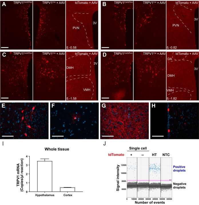
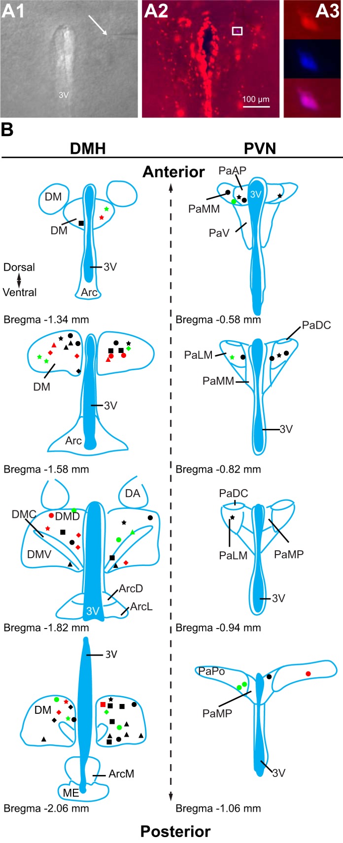
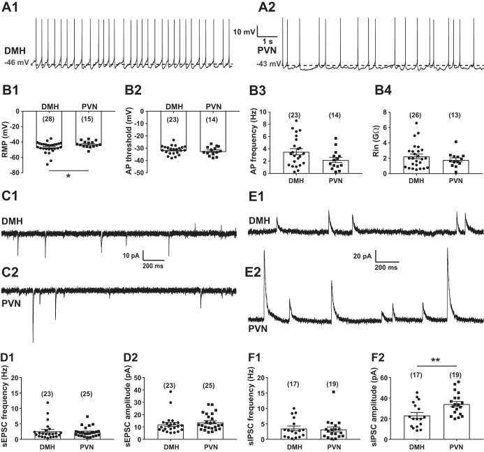
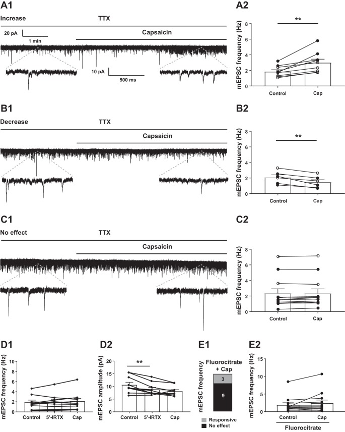
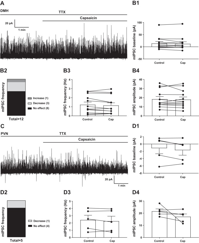
Transient receptor potential vanilloid type 1 (TRPV1) is a ligand-gated ion channel expressed in the peripheral and central nervous systems. TRPV1-dependent mechanisms take part in a wide range of physiological and pathophysiological pathways including the regulation of homeostatic functions. TRPV1 expression in the hypothalamus has been described as well as evidence that TRPV1-dependent excitatory inputs to hypothalamic preautonomic neurons are diminished in diabetic conditions. Here we aimed to determine the functional expression of TRPV1 in two hypothalamic nuclei known to be involved in the central control of metabolism and to test the hypothesis that TRPV1-expressing neurons receive TRPV1-expressing inputs. A mouse model (TRPV1Cre/tdTom) was generated to identify TRPV1-expressing cells and determine the cellular properties of TRPV1-expressing neurons in adult mice. Our study demonstrated the functional expression of TRPV1 in the dorsomedial hypothalamic nucleus and paraventricular nucleus in adult mice. Our findings revealed that a subset of TRPV1Cre/tdTom neurons receive TRPV1-expressing excitatory inputs, indicating direct interaction between TRPV1-expressing neurons. In addition, astrocytes likely play a role in the modulation of TRPV1-expressing neurons. In summary, this study identified specific hypothalamic regions where TRPV1 is expressed and functional in adult mice and the existence of direct connections between TRPV1Cre/tdTom neurons. NEW & NOTEWORTHY Transient receptor potential vanilloid type 1 (TRPV1) is expressed in the hypothalamus, and TRPV1-dependent regulation of preautonomic neurons is decreased in hyperglycemic conditions. Our study demonstrated functional expression of TRPV1 in two hypothalamic nuclei involved in the control of energy homeostasis. Our results also revealed that a subset of TRPV1-expressing neurons receive TRPV1-expressing excitatory inputs. These findings suggest direct interaction between TRPV1-expressing neurons.

Keywords: TRPV1; capsaicin; dorsomedial hypothalamic nucleus; paraventricular nucleus of the hypothalamus; patch-clamp.


Figures

Fig. 1.
Fig. 1.
Expression of transient receptor potential vanilloid type 1 (TRPV1) in the dorsomedial hypothalamic nucleus (DMH) and the paraventricular nucleus of the hypothalamus (PVN). A–D: representative images demonstrate TRPV1-expressing neurons in PVN (A and B) and DMH (C and D) in TRPV1Cre/tdTom mice (left), in TRPV1Cre mice injected with a Cre-dependent adeno-associated virus (AAV) construct (center), and in control tdTomato mice injected with the same viral construct (right). E–H: representative images show TRPV1 expression in neurons (E) and astrocyte-like cells (F) in the DMH, dense processes in the ventromedial hypothalamus (G), and ependymal-like cells lining the third ventricle (H). I: TRPV1 mRNA in the hypothalamus and cortex in adult mice. J: expression of TRPV1 in single cells. Droplet digital PCR was used to reveal TRPV1 expression in single cells. Positive droplets containing a single copy of TRPV1 cDNA (blue dots) were identified in tdTomato-expressing neurons and in diluted hypothalamic mRNA (HT; positive control), which demonstrates TRPV1 expression in tdTomato fluorescent neurons. There was no positive droplet in tdTomato-negative neurons or in the no-template control (NTC; negative control). Negative droplets (black dots) are droplets that do not contain TRPV1 cDNA. DA, dorsal hypothalamic area; VMH, ventromedial hypothalamic nucleus; 3V, 3rd ventricle. β, distance from bregma; tdTomato +, tdTomato-positive neuron; tdTomato, nonfluorescent neuron. Scale bars: 100 µm (A–D), 50 µm (E–H).
Fig. 2.
Fig. 2.
Location of recorded transient receptor potential vanilloid type 1 (TRPV1)-expressing neurons. A: patch-clamp recordings were conducted from TRPV1-expressing neurons. A1: differential interference contrast image during patch-clamp recording (×4). Arrow points to the tip of a recording pipette. A2: TRPV1-expressing neurons in the paraventricular nucleus of the hypothalamus (PVN; red, ×10). A3: enlarged view of boxed area shown in A2 (×40) illustrates a recorded TRPV1-expressing neuron (red) filled with Neurobiotin 350 (blue). B: schematic illustrations of the recorded TRPV1Cre/tdTom neurons in the dorsomedial hypothalamic nucleus (DMH) and the PVN. Each shape represents a different experimental setting as follows. In male mice: circle, miniature excitatory postsynaptic currents (mEPSCs) + capsaicin (Cap); square, mEPSCs +5′-iodoresiniferatoxin (5′-IRTX) + Cap; triangle, mEPSC + fluorocitrate (FC) + Cap; star, mIPSCs + Cap; in female mice: diamond, mEPSCs + Cap. Red indicates the location of neurons that had increased frequency after Cap application, green indicates the location of neurons that had decreased frequency, and black indicates the location of neurons not responding to Cap. 3V, 3rd ventricle; DM, DMH; DMD, dorsal part of DMH; DMC, compact part of DMH; DMV, ventral part of DMH; Arc, arcuate nucleus; ArcD, dorsal part of Arc; ArcL, lateral part of Arc; ArcM, medial part of Arc; ME, median eminence; DA, dorsal hypothalamic area; Pa, PVN; PaAP, anterior PVN; PaMM, medial magnocellular PVN; PaV, ventral PVN; PaDC, dorsal cap of PVN; PaLM, lateral PVN; PaMP, medial PVN; PaPO, posterior PVN. Schematics are based on Franklin and Paxinos (2007).
Fig. 3.
Fig. 3.
Basic cellular properties of transient receptor potential vanilloid type 1 (TRPV1)-expressing neurons in the dorsomedial hypothalamic nucleus (DMH) and the paraventricular nucleus of the hypothalamus (PVN). A: representative recordings from TRPV1-expressing neurons in the DMH (A1) and the PVN (A2). B: cellular properties of TRPV1-expressing neurons in the DMH and the PVN. B1: resting membrane potential (RMP). B2: threshold of action potentials (APs). B3: frequency of APs. B4: input resistance (Rin). C: representative recordings of spontaneous excitatory postsynaptic currents (sEPSCs) at holding potential of −60 mV in the DMH (C1) and the PVN (C2). D: frequency (D1) and amplitude (D2) of sEPSCs in TRPV1Cre/tdTom neurons in the DMH and the PVN. E: representative recordings of spontaneous inhibitory postsynaptic currents (sIPSCs) at holding potential of 0 mV in the DMH (E1) and the PVN (E2). F: frequency (F1) and amplitude (F2) of sIPSCs in TRPV1Cre/tdTom neurons in the DMH and the PVN. Numbers of recorded cells are shown in parentheses. *Significance (P < 0.05); **significance (P < 0.01).
Fig. 4.
Fig. 4.
Capsaicin (Cap) modulates excitatory neurotransmission of transient receptor potential vanilloid type 1 (TRPV1)-expressing neurons. A–C: Cap increased (A), decreased (B), or did not affect (C) the frequency of miniature excitatory postsynaptic currents (mEPSCs) in TRPV1Cre/tdTom neurons in the dorsomedial hypothalamic nucleus (DMH). Representative recordings of mEPSCs from TRPV1Cre/tdTom neurons in the DMH show increase (A1), decrease (B1), or no response (C1) to Cap application. TTX, tetrodotoxin. Bar graphs demonstrate increase (A2), decrease (B2), and no response (C2). D: in the presence of a TRPV1 antagonist, 5′-iodoresiniferatoxin (5′-IRTX), Cap did not increase mEPSC frequency (D1) and amplitude (D2); however, 5′-IRTX alone decreased mEPSC amplitude. E: in the presence of fluorocitrate, Cap increased mEPSC frequency in a small subset of TRPV1-expressing DMH neurons (E1), without altering the overall mEPSC frequency (E2). Numbers indicate number of neurons. ○, recording was conducted in female mice; ●, recording was conducted in male mice. **Significance (P < 0.01).
Fig. 5.
Fig. 5.
Capsaicin (Cap) does not modulate the overall inhibitory neurotransmission of TRPV1Cre/tdTom neurons. A: representative recording of miniature inhibitory postsynaptic currents (mIPSCs) from a transient receptor potential vanilloid type 1 (TRPV1)-expressing dorsomedial hypothalamic nucleus (DMH) neuron. TTX, tetrodotoxin. B: Cap had no significant effect on the baseline (B1), overall frequency (B3), and amplitude (B4) of mIPSCs in TRPV1Cre/tdTom DMH neurons. C: representative recording of mIPSCs from a TRPV1-expressing paraventricular nucleus of the hypothalamus (PVN) neuron. D: Cap had no significant effect on the baseline (D1), overall frequency (D3), and amplitude (D4) of mIPSCs in TRPV1Cre/tdTom PVN neurons. Numbers in B2 and D2 indicate number of neurons.

References

    1. Akiba Y, Kato S, Katsube K, Nakamura M, Takeuchi K, Ishii H, Hibi T. Transient receptor potential vanilloid subfamily 1 expressed in pancreatic islet beta cells modulates insulin secretion in rats. Biochem Biophys Res Commun 321: 219–225, 2004. doi:10.1016/j.bbrc.2004.06.149. - DOI - PubMed
    1. Baboota RK, Singh DP, Sarma SM, Kaur J, Sandhir R, Boparai RK, Kondepudi KK, Bishnoi M. Capsaicin induces “brite” phenotype in differentiating 3T3-L1 preadipocytes. PLoS One 9: e103093, 2014. doi:10.1371/journal.pone.0103093. - DOI - PMC - PubMed
    1. Bari M, Bonifacino T, Milanese M, Spagnuolo P, Zappettini S, Battista N, Giribaldi F, Usai C, Bonanno G, Maccarrone M. The endocannabinoid system in rat gliosomes and its role in the modulation of glutamate release. Cell Mol Life Sci 68: 833–845, 2011. doi:10.1007/s00018-010-0494-4. - DOI - PMC - PubMed
    1. Boychuk CR, Zsombok A, Tasker JG, Smith BN. Rapid glucocorticoid-induced activation of TRP and CB1 receptors causes biphasic modulation of glutamate release in gastric-related hypothalamic preautonomic neurons. Front Neurosci 7: 3, 2013. doi:10.3389/fnins.2013.00003. - DOI - PMC - PubMed
    1. Caterina MJ, Schumacher MA, Tominaga M, Rosen TA, Levine JD, Julius D. The capsaicin receptor: a heat-activated ion channel in the pain pathway. Nature 389: 816–824, 1997. doi:10.1038/39807. - DOI - PubMed

Publication types

MeSH terms

LinkOut - more resources