. 2019 Jan 10;47(1):3-14.
doi: 10.1093/nar/gky1163.

Inosine induces context-dependent recoding and translational stalling

Affiliations

Inosine induces context-dependent recoding and translational stalling

Konstantin Licht et al. Nucleic Acids Res. .

Abstract

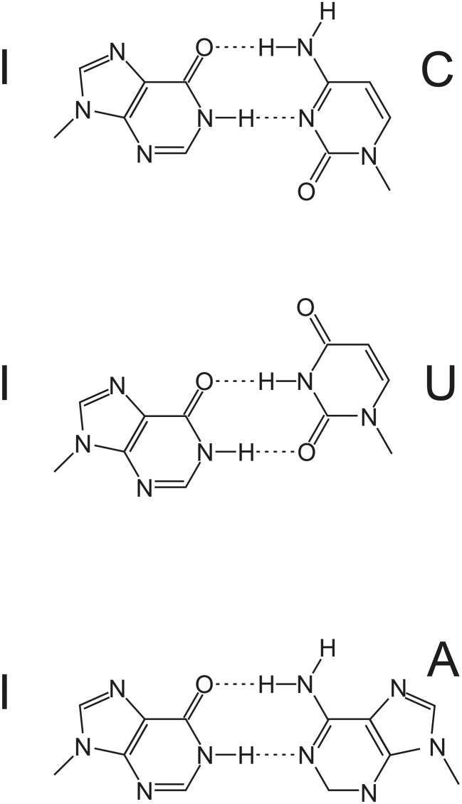
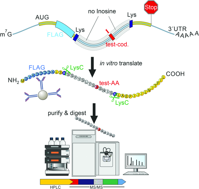
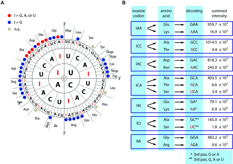
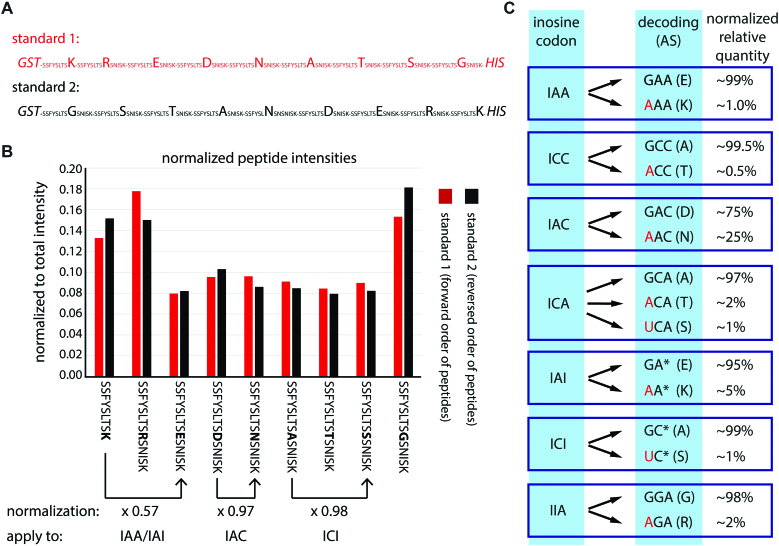
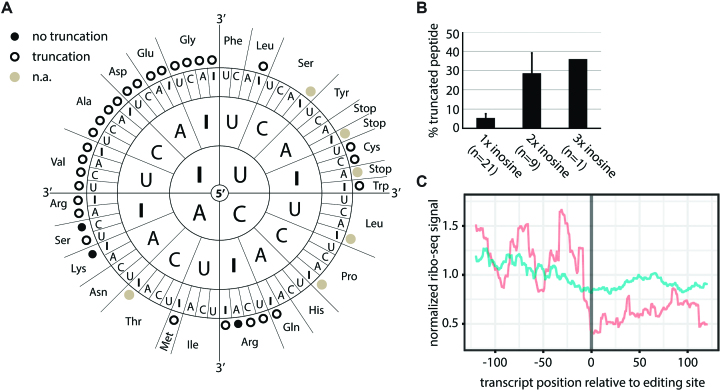
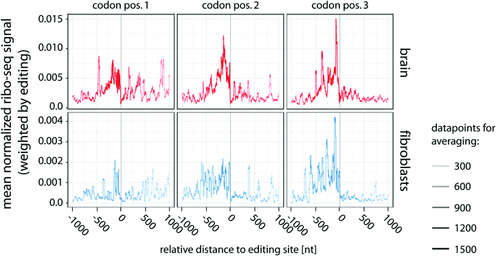
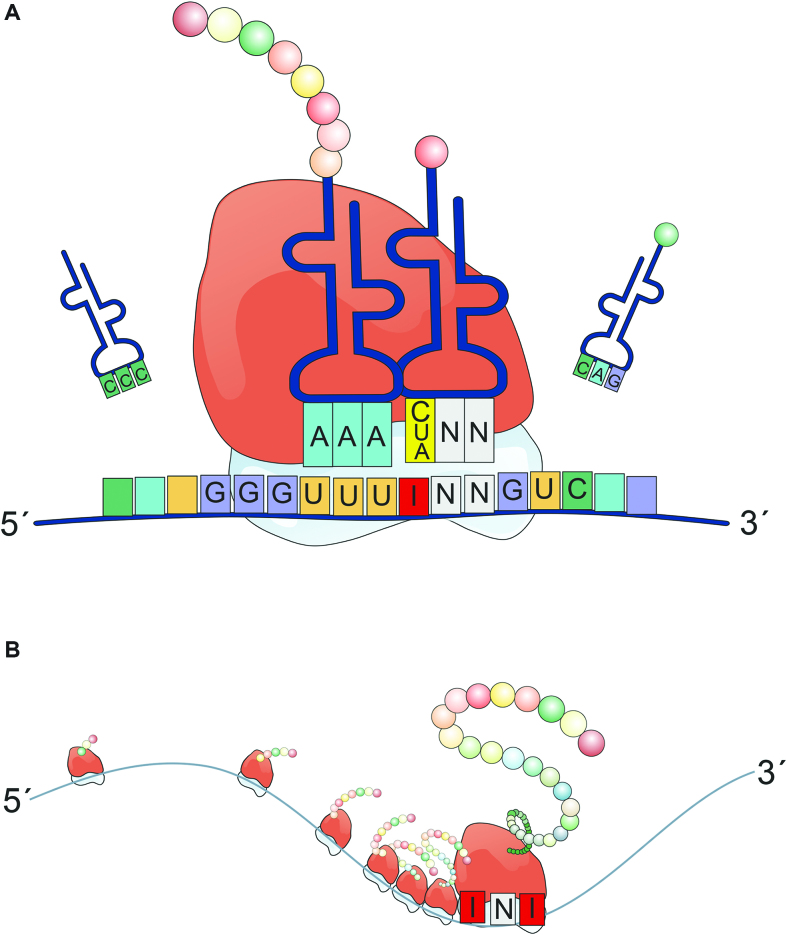
RNA modifications are present in all classes of RNAs. They control the fate of mRNAs by affecting their processing, translation, or stability. Inosine is a particularly widespread modification in metazoan mRNA arising from deamination of adenosine catalyzed by the RNA-targeting adenosine deaminases ADAR1 or ADAR2. Inosine is commonly thought to be interpreted as guanosine by cellular machines and during translation. Here, we systematically test ribosomal decoding using mass spectrometry. We show that while inosine is primarily interpreted as guanosine it can also be decoded as adenosine, and rarely even as uracil. Decoding of inosine as adenosine and uracil is context-dependent. In addition, mass spectrometry analysis indicates that inosine causes ribosome stalling especially when multiple inosines are present in the codon. Indeed, ribosome profiling data from human tissues confirm inosine-dependent ribosome stalling in vivo. To our knowledge this is the first study where decoding of inosine is tested in a comprehensive and unbiased way. Thus, our study shows novel, unanticipated functions for inosines in mRNAs, further expanding coding potential and affecting translational efficiency.


Figures

Figure 1.
Figure 1.
Inosine can basepair with cytidine, uracil or adenine, in each case forming two hydrogen bonds.
Figure 2.
Figure 2.
A setup to systematically test for the decoding of inosines in mRNA. A DNA template was designed that allows the in vitro transcription of an RNA containing a single inosine containing codon (red). The RNA also encodes a FLAG tag (light blue) and two lysine codons (dark blue). Upon in vitro translation using rabbit reticulocyte lysate the short protein can be purified using anti-FLAG antibodies. Following cleavage of the translated protein with LysC the peptide containing a single test amino acid (AA, red) is identified by mass spectrometry.
Figure 3.
Figure 3.
Context-dependent decoding of inosines as guanosine, adenosine, or uracil. (A) Scheme showing all codons where substitution of G by I may lead to recoding. For multiple codons peptide variants were detected that support decoding of inosine as adenosine or uracil (I = G, A, or U; A or U >0.4%; red dots). Codons giving rise to ambiguous products due to redundancy of the genetic code were omitted (n.a.; gray dots). Codons where only guanosine-decoding was detected are marked by a blue dot. (B) The peak intensities of the detected peptides supporting primary and alternative decoding are given (see Supplementary Table S1 for all tested codons). Alternative decoding is highlighted in red. Two test-codons contain inosine in the wobble position (marked by * or **). As either *A/G or **A/G/U would result in the same amino acid we cannot make a statement of the decoding at this position.
Figure 4.
Figure 4.
Alternative decoding is significant for codons IAC and IAI. (A) For normalization of mass spectrometry results, all peptides with substantial alternative decoding were expressed as one concatenated peptide in two orders (forward: red, reverse: black) in E. coli with an N-terminal GST-tag and a C-terminal His-tag. The peptide sequence is given and changed amino acids are highlighted. (B) After expression and purification the individual peptides were released using Lys-C and measured by mass spectrometry. The individual peptide intensities were normalized to the summed total intensity. The peptide sequence is given below. For differences in intensity between two peptides a normalization factor (normalization) is calculated based on the average intensity between standard 1 and standard 2. Three examples are given below the peptide sequence. The factors are then applied to the peptide intensities detected after in vitro translation shown in: (C) The relative primary and alternative decoding after normalization are given. Red indicates alternative decoding. The detected amino acid is shown in brackets.
Figure 5.
Figure 5.
Inosine causes ribosome stalling. (A) Black dots indicate exclusive detection of full-length peptide, circles indicate additional truncated peptides. Codons with inosine only in the wobble position or STOP codons were omitted (n.a.; gray dots). (B) Percentage of truncated peptide detected for different numbers of inosines in the codon are shown. Error bars = s.e.m. (C) Inosine levels at known editing sites were calculated from brain mRNA-seq data. Ribosome profiling data for sites showing editing (red) or no editing (blue) were normalized, weighted by editing rate, merged, and centered on the editing site. The coverage from position −125 to +125 relative to the editing site is given.
Figure 6.
Figure 6.
Inosine-induced ribosome stalling is position-independent and occurs in multiple tissues. Inosine levels at known editing sites were calculated from brain mRNA-seq data (top). Ribosome profiling data (ribo-seq) were normalized, weighted by editing rate, merged, and centered on the editing site split according to the position of the inosine in the codon (codon pos.1–3). The coverage from position −1000 to +1000 relative−to the editing site is given. For comparison, ribosome-profiling data from interferon-induced fibroblasts was analyzed (bottom). The number of data points used for averaging is reflected by the thickness of the line.
Figure 7.
Figure 7.
Inosines in mRNAs can lead to different recoding events and promote ribosome stalling. (A) A single inosine in a codon of an mRNA can basepair with C, U, or A in the corresponding tRNA, leading to different decoding events. (B) Especially the presence of multiple inosines but also individual inosines in a codon seemingly provokes ribosome stalling and the formation of truncated peptides.

References

    1. Machnicka M.A., Milanowska K., Osman Oglou O., Purta E., Kurkowska M., Olchowik A., Januszewski W., Kalinowski S., Dunin-Horkawicz S., Rother K.M. et al. . MODOMICS: a database of RNA modification pathways–2013 update. Nucleic Acids Res. 2013; 41:D262–D267. - PMC - PubMed
    1. Licht K., Jantsch M.F.. Rapid and dynamic transcriptome regulation by RNA editing and RNA modifications. J. Cell Biol. 2016; 213:15–22. - PMC - PubMed
    1. Zhao B.S., Roundtree I.A., He C.. Post-transcriptional gene regulation by mRNA modifications. Nat. Rev. Mol. Cell Biol. 2017; 18:31–42. - PMC - PubMed
    1. Huang Y.S., Lu W.H.. Decoding hidden messages in neurons: insights from epitranscriptome-controlled and specialized ribosome-controlled translation. Curr. Opin. Neurobiol. 2018; 48:64–70. - PubMed
    1. Saletore Y., Meyer K., Korlach J., Vilfan I.D., Jaffrey S., Mason C.E.. The birth of the Epitranscriptome: deciphering the function of RNA modifications. Genome Biol. 2012; 13:175. - PMC - PubMed

Publication types

MeSH terms