

Pilot study for three-dimensional assessment of laminar pore structure in patients with glaucoma, as measured with swept source optical coherence tomography
- PMID: 30462712
- PMCID: PMC6248986
- DOI: 10.1371/journal.pone.0207600
Pilot study for three-dimensional assessment of laminar pore structure in patients with glaucoma, as measured with swept source optical coherence tomography
Abstract
Purpose: To develop a method to quantify, based on swept-source optical coherence tomography (OCT), the 3D structure of the laminar pores in patients with glaucoma.
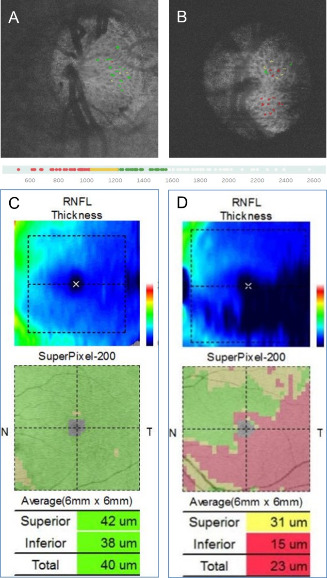
Methods: This retrospective study examined 160 laminar pores from 8 eyes of 8 cases: 4 normal subjects and 4 open-angle glaucoma (OAG) patients. We reconstructed 3D volume data for a 3 x 3 mm disc, using a method similar to OCT angiography, and segmented the structure of the lamina cribrosa. Then, we manually segmented each laminar pore in sequential C-scan images (>90 slices at 2.6-micron intervals) with VCAT5 (RIKEN, Japan). We compared the control and OAG subjects with the Mann-Whitney U test. Differences were considered significant at p < 0.05.
Results: We found that the laminar pores of the OAG patients had a significantly smaller average cross-sectional area, smaller 3D volume (adjusted to the average thickness of the lamina cribrosa), and higher true sphericity, and lower principal value (P1, 2, 3) of the 3D structure data (all: p < 0.0001). The topographic distribution of damaged laminar pores was consistent with the damaged area of the macular map.
Conclusion: We successfully developed a method to quantify the 3D structure of the laminar pores; providing a useful tool to assess lamina cribrosa-associated risk factors for glaucoma. These findings promise to benefit future investigations into the pathomechanisms of glaucoma.
Conflict of interest statement
Co-authors GA, TK, and MA are employed by Topcon Corporation, a commercial company. There are no patents, products in development or marketed products to declare. This does not alter our adherence to all the PLOS ONE policies on sharing data and materials.
Figures


Similar articles
-
Three-dimensional high-speed optical coherence tomography imaging of lamina cribrosa in glaucoma.Ophthalmology. 2009 Feb;116(2):214-22. doi: 10.1016/j.ophtha.2008.09.008. Epub 2008 Dec 16. Ophthalmology. 2009. PMID: 19091413
-
Three-dimensional imaging of lamina cribrosa defects in glaucoma using swept-source optical coherence tomography.Invest Ophthalmol Vis Sci. 2013 Jul 18;54(7):4798-807. doi: 10.1167/iovs.13-11677. Invest Ophthalmol Vis Sci. 2013. PMID: 23778878
-
Imaging the posterior segment of the eye using swept-source optical coherence tomography in myopic glaucoma eyes: comparison with enhanced-depth imaging.Am J Ophthalmol. 2014 Mar;157(3):550-7. doi: 10.1016/j.ajo.2013.11.008. Epub 2013 Nov 12. Am J Ophthalmol. 2014. PMID: 24239773
-
[Aiming for zero blindness].Nippon Ganka Gakkai Zasshi. 2015 Mar;119(3):168-93; discussion 194. Nippon Ganka Gakkai Zasshi. 2015. PMID: 25854109 Review. Japanese.
-
Improved visualization of deep ocular structures in glaucoma using high penetration optical coherence tomography.Expert Rev Med Devices. 2013 Sep;10(5):621-8. doi: 10.1586/17434440.2013.827505. Epub 2013 Aug 23. Expert Rev Med Devices. 2013. PMID: 23972075 Review.
Cited by
-
Morphological Changes of Glial Lamina Cribrosa of Rats Suffering from Chronic High Intraocular Pressure.Bioengineering (Basel). 2022 Nov 30;9(12):741. doi: 10.3390/bioengineering9120741. Bioengineering (Basel). 2022. PMID: 36550947 Free PMC article.
-
Lamina Cribrosa Microstructure in Nonhuman Primates With Naturally Occurring Peripapillary Retinal Nerve Fiber Layer Thinning.Transl Vis Sci Technol. 2024 Sep 3;13(9):23. doi: 10.1167/tvst.13.9.23. Transl Vis Sci Technol. 2024. PMID: 39297808 Free PMC article.
References
-
- Leske MC, Heijl A, Hyman L, Bengtsson B, Dong L, Yang Z, et al. Predictors of long-term progression in the early manifest glaucoma trial. Ophthalmology. 2007;114(11):1965–72. 10.1016/j.ophtha.2007.03.016 - DOI - PubMed
-
- Suzuki Y, Iwase A, Araie M, Yamamoto T, Abe H, Shirato S, et al. Risk factors for open-angle glaucoma in a Japanese population: the Tajimi Study. Ophthalmology. 2006;113(9):1613–7. 10.1016/j.ophtha.2006.03.059 - DOI - PubMed
-
- Marcus MW, de Vries MM, Junoy Montolio FG, Jansonius NM. Myopia as a risk factor for open-angle glaucoma: a systematic review and meta-analysis. Ophthalmology. 2011;118(10):1989–94 e2. 10.1016/j.ophtha.2011.03.012 - DOI - PubMed
-
- Tielsch JM, Katz J, Sommer A, Quigley HA, Javitt JC. Family history and risk of primary open angle glaucoma. The Baltimore Eye Survey. Arch Ophthalmol. 1994;112(1):69–73. - PubMed
-
- Faridi OS, Park SC, Kabadi R, Su D, De Moraes CG, Liebmann JM, et al. Effect of focal lamina cribrosa defect on glaucomatous visual field progression. Ophthalmology. 2014;121(8):1524–30. 10.1016/j.ophtha.2014.02.017 - DOI - PubMed
Publication types
MeSH terms
LinkOut - more resources
Full Text Sources
