

3T magnetic resonance spectroscopy as a powerful diagnostic modality for assessment of thyroid nodules
- PMID: 30462802
- PMCID: PMC10118653
- DOI: 10.20945/2359-3997000000069
3T magnetic resonance spectroscopy as a powerful diagnostic modality for assessment of thyroid nodules
Abstract
Objective: Magnetic resonance spectroscopy (MRS) is a powerful tool for structural studies of chemical compounds and biomolecules and also documented promising findings as a potential imaging technology in thyroid oncology. This prospective study was to ascertain the clinical significance of 3 Tesla MRS in the evaluation of patients with thyroid nodules (TNs) as an ancillary diagnostic technique for thyroid carcinoma.
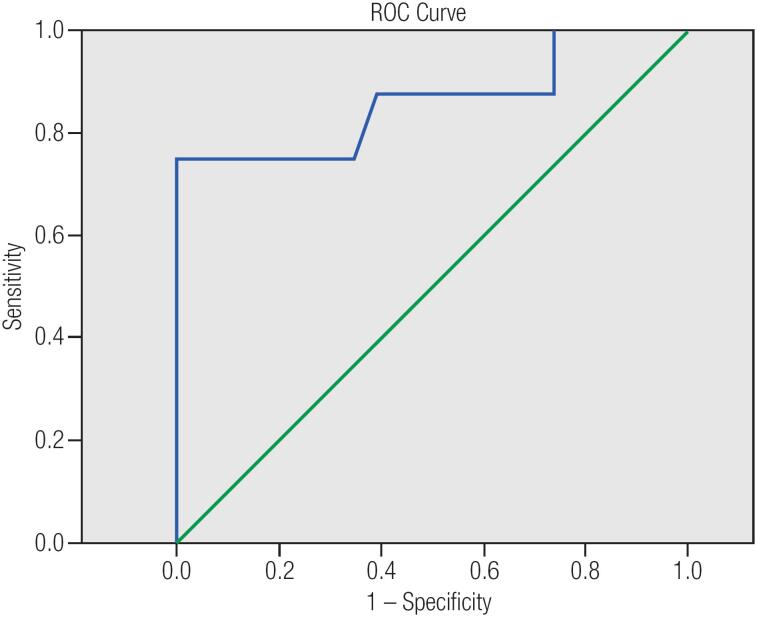
Materials and methods: Magnetic resonance spectroscopy at 3T at echo- times (TEs) 136 and 270 ms was carried out on 15 patients with total number of 32 TNs larger than 1 cm3, which all were surgically resected. Choline (Chol) to creatine (Cr) ratio was assessed at 136 and 270 TEs on each nodule and a receiver operating characteristic (ROC) curve was used to determine optimal cut-off point. The findings were compared with histopathology of thyroid specimens.
Results: There were 23 benign and 9 malignant lesions (7 papillary and 2 follicular thyroid carcinomas). The mean values of Chol/Cr at 136 and 270 TEs was 2.28 ± 3.65 and 1.52 ± 1.67 respectively and the difference between benign and malignant nodules was only significant at 136 TEs. The study revealed that Chol/ Cr ratio cut-off point of 2.5 best correlates with histopathology results (sensitivity = 75%; specificity = 100%; PPV = 100%; NPV= 92%).
Conclusion: This preliminary study showed that 3T magnetic resonance spectroscopy might be a specific modality for the evaluation of thyroid nodules in differentiation of benign from malignant thyroid tissue. However, a larger series would give much greater confidence that this state-of-the-art technology will worth pursuing in clinical practice.
Figures

Similar articles
-
T2* mapping at 3.0T MRI for differentiation of papillary thyroid carcinoma from benign thyroid nodules.J Magn Reson Imaging. 2016 Apr;43(4):956-61. doi: 10.1002/jmri.25041. Epub 2015 Sep 21. J Magn Reson Imaging. 2016. PMID: 26389559
-
The role of proton MR spectroscopy and apparent diffusion coefficient values in the diagnosis of malignant thyroid nodules: preliminary results.Clin Imaging. 2012 Jul-Aug;36(4):323-33. doi: 10.1016/j.clinimag.2011.09.009. Epub 2012 Jun 8. Clin Imaging. 2012. PMID: 22726971
-
Magnetic resonance spectroscopy as a diagnostic modality for carcinoma thyroid.Eur J Radiol. 2007 Dec;64(3):414-8. doi: 10.1016/j.ejrad.2007.03.006. Epub 2007 Apr 26. Eur J Radiol. 2007. PMID: 17462842
-
Differentiation between malignant and benign thyroid nodules and stratification of papillary thyroid cancer with aggressive histological features: Whole-lesion diffusion-weighted imaging histogram analysis.J Magn Reson Imaging. 2016 Dec;44(6):1546-1555. doi: 10.1002/jmri.25290. Epub 2016 Apr 19. J Magn Reson Imaging. 2016. PMID: 27093648
-
Role of duplex power Doppler ultrasound in differentiation between malignant and benign thyroid nodules.Korean J Radiol. 2010 Nov-Dec;11(6):594-602. doi: 10.3348/kjr.2010.11.6.594. Epub 2010 Oct 29. Korean J Radiol. 2010. PMID: 21076584 Free PMC article.
Cited by
-
Malignant and benign thyroid nodule differentiation through the analysis of blood plasma with terahertz spectroscopy.Biomed Opt Express. 2021 Jan 26;12(2):1020-1035. doi: 10.1364/BOE.412715. eCollection 2021 Feb 1. Biomed Opt Express. 2021. PMID: 33680557 Free PMC article.
-
Metabolomics of thyroid nodules and the future.Arch Endocrinol Metab. 2018 Oct;62(5):493-494. doi: 10.20945/2359-3997000000080. Arch Endocrinol Metab. 2018. PMID: 30462800 Free PMC article. No abstract available.
References
-
- Burman KD, Wartofsky L. Clinical Practice. Thyroid Nodules. N Engl J Med. 2015;373(24):2347-56. - PubMed
-
- Durante C, Costante G, Lucisano G, Bruno R, Meringolo D, Paciaroni A, et al. The natural history of benign thyroid nodules. JAMA. 2015;313(9):926-35. - PubMed
-
- Mandell DL, Genden EM, Mechanick JI, Bergman DA, Biller HF, Urken ML. Diagnostic accuracy of fine-needle aspiration and frozen section in nodular thyroid disease. Otolaryngol Head Neck Surg. 2001;124(5):531-6. - PubMed
-
- Riazi A, Kalantarhormozi M, Nabipour I, Eghbali SS, Farzaneh M, Javadi H, et al. Technetium-99m methoxyisobutylisonitrile scintigraphy in the assessment of cold thyroid nodules: is it time to change the approach to the management of cold thyroid nodules? Nucl Med Commun. 2014;35(1):51-7. - PubMed
-
- Riazi A, Kalantarhormozi M, Nabipour I, Ostovar A, Javadi H, Assadi M. Comments on Wale et al.: Combined (99)mTc-methoxyisobutylisonitrile scintigraphy and fine-needle aspiration cytology offers an accurate and potentially cost-effective investigative strategy for the assessment of solitary or dominant thyroid nodules. Eur J Nucl Med Mol Imaging. 2014;41(3):575-6. - PubMed
Publication types
MeSH terms
Substances
LinkOut - more resources
Full Text Sources
Medical