Review
. 2018 Nov 20;19(11):3672.
doi: 10.3390/ijms19113672.

Epithelial-Mesenchymal Transition and Metastasis under the Control of Transforming Growth Factor β

Affiliations
Review

Epithelial-Mesenchymal Transition and Metastasis under the Control of Transforming Growth Factor β

Yutaro Tsubakihara et al. Int J Mol Sci. .

Abstract

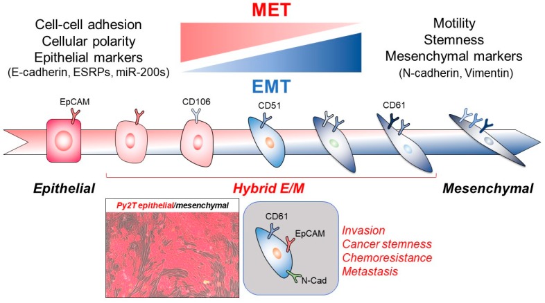
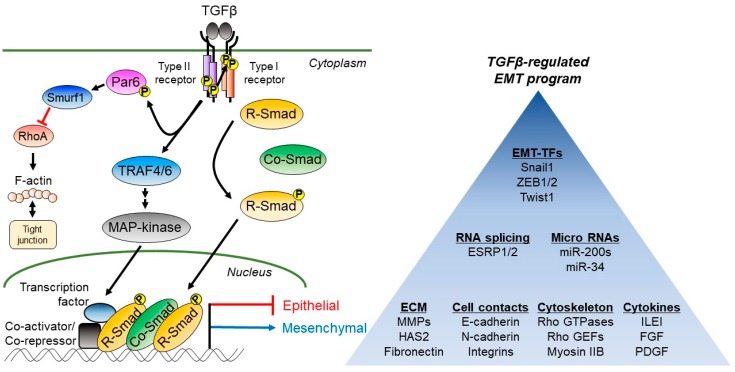
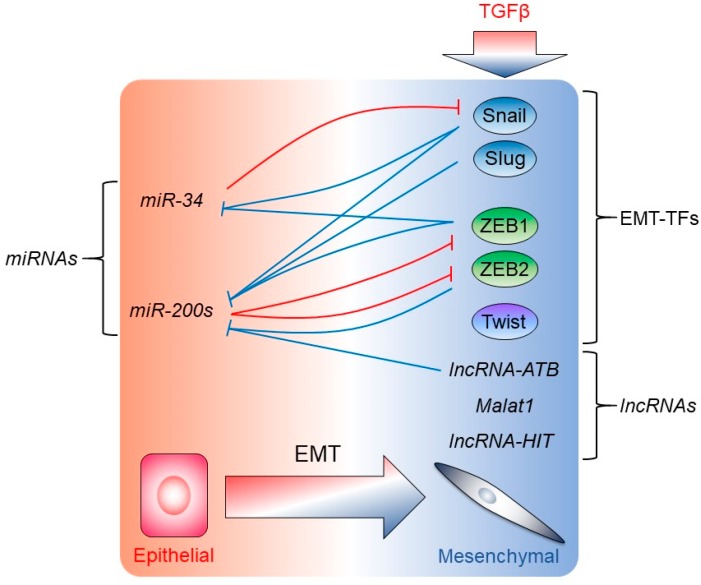
Metastasis of tumor cells from primary sites of malignancy to neighboring stromal tissue or distant localities entails in several instances, but not in every case, the epithelial-mesenchymal transition (EMT). EMT weakens the strong adhesion forces between differentiated epithelial cells so that carcinoma cells can achieve solitary or collective motility, which makes the EMT an intuitive mechanism for the initiation of tumor metastasis. EMT initiates after primary oncogenic events lead to secondary secretion of cytokines. The interaction between tumor-secreted cytokines and oncogenic stimuli facilitates EMT progression. A classic case of this mechanism is the cooperation between oncogenic Ras and the transforming growth factor β (TGFβ). The power of TGFβ to mediate EMT during metastasis depends on versatile signaling crosstalk and on the regulation of successive waves of expression of many other cytokines and the progressive remodeling of the extracellular matrix that facilitates motility through basement membranes. Since metastasis involves many organs in the body, whereas EMT affects carcinoma cell differentiation locally, it has frequently been debated whether EMT truly contributes to metastasis. Despite controversies, studies of circulating tumor cells, studies of acquired chemoresistance by metastatic cells, and several (but not all) metastatic animal models, support a link between EMT and metastasis, with TGFβ, often being a common denominator in this link. This article aims at discussing mechanistic cases where TGFβ signaling and EMT facilitate tumor cell dissemination.

Keywords: epithelial-mesenchymal transition; micro-RNA; non-coding RNA; signal transduction; transcription factor; transforming growth factor β; tumor invasiveness.


Conflict of interest statement

The authors declare no conflict of interest.

Figures

Figure 1
Figure 1
The trajectory of differentiation change is pictorially graphed with epithelial cells on the left, hybrid E/M cells in the middle, and mesenchymal cells on the right hand of the arrow. The EMT and MET are also depicted as gradients of molecular and phenotypic change at the top of the figure and inside the main trajectory. Specific molecular and cellular attributes of epithelial and mesenchymal cells are listed on top of the relevant cell types. Important cell surface antigens are also drawn on the plasma membrane of each cell in a different color, in order to mark the molecular progression from an epithelial to a mesenchymal phenotype and the intermediate stages. At the bottom, the photomicrograph shows a mixed population of epithelial (red) and mesenchymal (dark grey) Py2T mouse breast cancer cells that have undergone EMT followed by MET. An additional cell model depicts a possible hybrid E/M cell that expresses EpCAM, CD61, and N-cadherin (N-Cad) as revealed in certain studies of circulating tumor cells. The features of EMT that are relevant to cancer are listed on the right.
Figure 2
Figure 2
Basic TGFβ signaling diagram along with a program of TGFβ-regulated genes that contribute to the EMT. Left side, the extracellular dimeric TGFβ ligand is shown to bind to its plasma membrane receptors, the type II and type I receptors (each drawn as a dimer), causing trans-phosphorylation (circled P) of the type I receptor by the type II receptor. The type II receptor phosphorylates the polarity protein Par6, which recruits the ubiquitin ligase Smurf1 and regulates RhoA-dependent actin assembly and tight junction disassembly. The type I receptor also recruits the ubiquitin ligases TRAF4 and TRAF6, which activate the MAP-kinase pathway by ubiquitination, leading to the transcription factor phosphorylation. The type I receptor kinase also phosphorylates R-Smads, which form complexes with the Co-Smad, Smad4. In the nucleus, Smad complexes and cooperating transcription factors bound to various genes, along with co-repressors (co-activators) either repress the expression of epithelial genes or induce the expression of mesenchymal genes. Right side, a summary of the TGFβ-regulated EMT program divided into seven subprograms, each enlisting only a small representative example of genes that are involved in the EMT and cell motility. Signaling flow is indicated by black arrows (positive flow) and red T-bars (negative regulation); on the gene transcriptional start site, a red T-bar indicates negative regulation and a blue arrow indicates positive regulation of transcription.
Figure 3
Figure 3
The non-coding RNAs regulate the EMT. Epithelial (left) and mesenchymal (right) genes are listed, the latter being transcriptionally induced by TGFβ signaling (thick arrow). Thin T-bars indicate the negative regulation of EMT-TFs by miRNAs and inversely, the negative regulation of miRNA expression by the EMT-TFs or lncRNAs. Negative transcriptional regulation is shown with blue T-bars, whereas negative regulation of mRNA translation and stability by miRNAs is shown with red T-bars.

Similar articles

Cited by

References

    1. Nieto M.A., Huang R.Y., Jackson R.A., Thiery J.-P. EMT 2016. Cell. 2016;166:21–45. doi: 10.1016/j.cell.2016.06.028. - DOI - PubMed
    1. Kahata K., Dadras M.S., Moustakas A. TGF-β family signaling in epithelial differentiation and epithelial-mesenchymal transition. Cold Spring Harb. Perspect. Biol. 2018;10 doi: 10.1101/cshperspect.a022194. - DOI - PMC - PubMed
    1. Ye X., Weinberg R.A. Epithelial-Mesenchymal Plasticity: A Central Regulator of Cancer Progression. Trends Cell Biol. 2015;25:675–686. doi: 10.1016/j.tcb.2015.07.012. - DOI - PMC - PubMed
    1. Gal A., Sjöblom T., Fedorova L., Imreh S., Beug H., Moustakas A. Sustained TGFβ exposure suppresses Smad and non-Smad signalling in mammary epithelial cells, leading to EMT and inhibition of growth arrest and apoptosis. Oncogene. 2008;27:1218–1230. doi: 10.1038/sj.onc.1210741. - DOI - PubMed
    1. Jolly M.K., Ware K.E., Gilja S., Somarelli J.A., Levine H. EMT and MET: Necessary or permissive for metastasis? Mol. Oncol. 2017;11:755–769. doi: 10.1002/1878-0261.12083. - DOI - PMC - PubMed

MeSH terms

Substances

LinkOut - more resources