

Performance of a bioglass-based dentine desensitizer under lactid acid exposition: an in-vitro study
- PMID: 30463552
- PMCID: PMC6249813
- DOI: 10.1186/s12903-018-0642-z
Performance of a bioglass-based dentine desensitizer under lactid acid exposition: an in-vitro study
Erratum in
-
Correction to: Performance of a bioglass-based dentine desensitizer under lactid acid exposition: an in-vitro study.BMC Oral Health. 2019 Jul 24;19(1):160. doi: 10.1186/s12903-019-0855-9. BMC Oral Health. 2019. PMID: 31340807 Free PMC article.
Abstract
Background: Dentine hypersensitivity is especially frequent in patients with pronounced periodontal attachment loss. Aim of the treatment is an obstruction of the dentine tubules in order to inhibit liquid or osmotic motion, which is considered as trigger for pain sensations. Novel approaches aim for obstruction by calcium phosphate compounds in order to rely on biocompatible compounds. It was the aim of the study to optically investigate the morphology and to assess the fluid permeability of treated dentine surfaces.
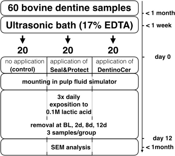
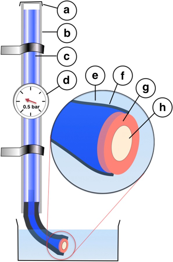
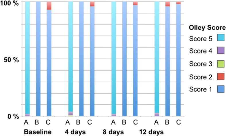
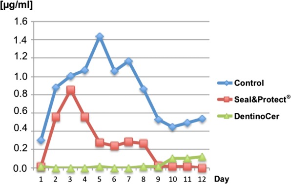
Methods: Dentine discs were pretreated in an ultrasonic bath with 17% EDTA to clean the lumina of the dentine tubules. Samples of group A remained untreated while Seal&Protect® as a conventional desensitizer was applied for group B and DentinoCer in group C. Discs were mounted into a pulp fluid simulator (PFS) with a methylene blue solution in order to create a flow pressure of 0.5 bar. Over 12 d, discs were exposed three times per day to 0.1 M nonsaturated lactic acid. At baseline and after 2, 8 and 12 d samples were removed from PFS and prepared for SEM analysis. Tubule obstruction was assessed quantitatively using Olley scores and by qualitative description of the surface. Absorption spectrometry was used to assess the concentration of leaked methylene blue outside the samples in order to estimate dentine permeability.
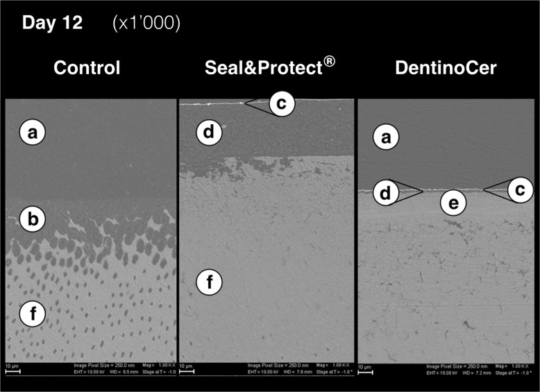
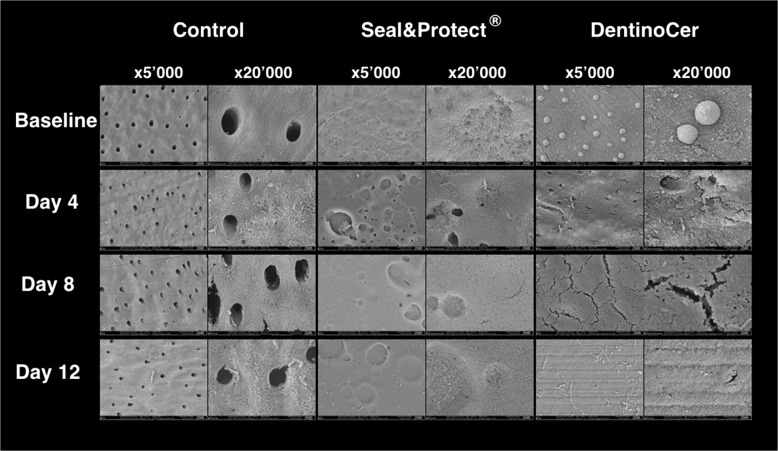
Results: Untreated discs showed clean lumina of all tubules at all time points and magnifications. From day 2 onwards dentine showed exposed collagene fibers due to acid exposition. Seal&Protect® initially showed homogenous dentine surface coverage that got a more granulomatous aspect in the course of treatment time. Few samples showed sporadic tubules with open lumen at day 8 and 12. Group C showed samples with a homogeneous, even surface. Narrow slits in the superficial layer are visible from day 4 on, but the dentine surface remained invisible and dentine tubules were closed till the end of the investigation period.
Conclusion: Over 12 d of lactid acid exposure, samples showed complete coverage of the dentine tubules in the chosen in-vitro-model when treated with Seal&Protect® or DentinoCer.
Keywords: Dentine sensitivity; Desensitizer; Electron microscopy; Hypersensitivity; Periodontitis.
Conflict of interest statement
Ethics approval and consent to participate
Since no human beings have been involved in this study, according to Swiss national and cantonal law an ethics approval is not needed and a consent to participate is not applicable. Bovine dentine discs were gained from jaws of slaughter cattle. No living animals have been interacting with the conduction of the present study.
Consent for publication
Not applicable.
Competing interests
The authors declare that they have no competing interests.
Publisher’s Note
Springer Nature remains neutral with regard to jurisdictional claims in published maps and institutional affiliations.
Figures
References
Publication types
MeSH terms
Substances
LinkOut - more resources
Full Text Sources
