. 2018 Nov 21;19(1):229.
doi: 10.1186/s12931-018-0938-1.

Alterations in VASP phosphorylation and profilin1 and cofilin1 expression in hyperoxic lung injury and BPD

Affiliations

Alterations in VASP phosphorylation and profilin1 and cofilin1 expression in hyperoxic lung injury and BPD

Mehboob Ali et al. Respir Res. .

Abstract

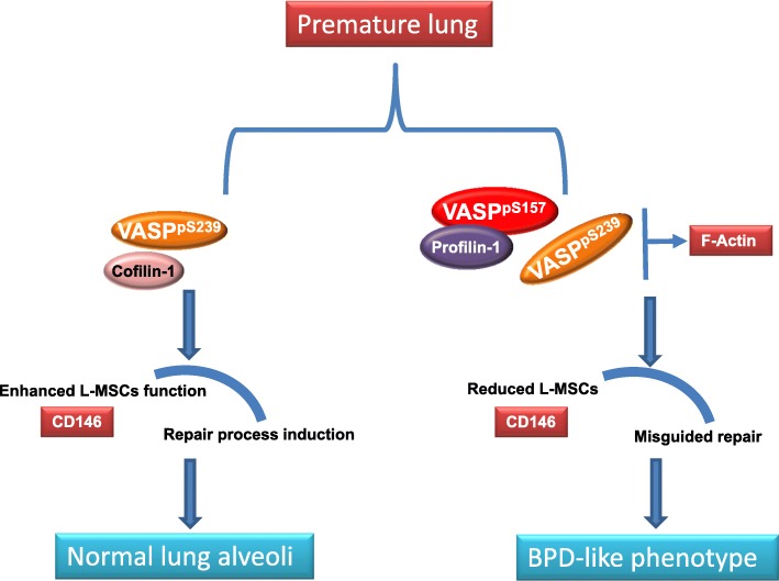
Background: Hyperoxia is a frequently employed therapy for prematurely born infants, induces lung injury and contributes to development of bronchopulmonary dysplasia (BPD). BPD is characterized by decreased cellular proliferation, cellular migration, and failure of injury repair systems. Actin binding proteins (ABPs) such as VASP, cofilin1, and profilin1 regulate cell proliferation and migration via modulation of actin dynamics. Lung mesenchymal stem cells (L-MSCs) initiate repair processes by proliferating, migrating, and localizing to sites of injury. These processes have not been extensively explored in hyperoxia induced lung injury and repair.

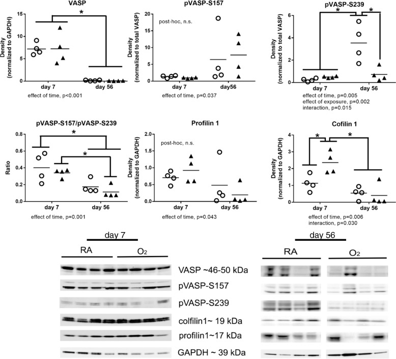
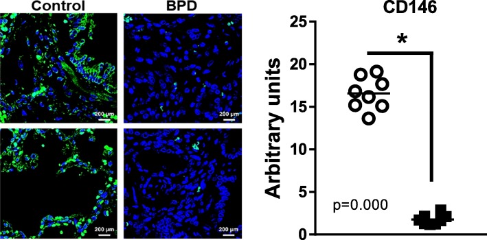
Methods: ABPs and CD146+ L-MSCs were analyzed by immunofluorescence in human lung autopsy tissues from infants with and without BPD and by western blot in lung tissue homogenates obtained from our murine model of newborn hyperoxic lung injury.

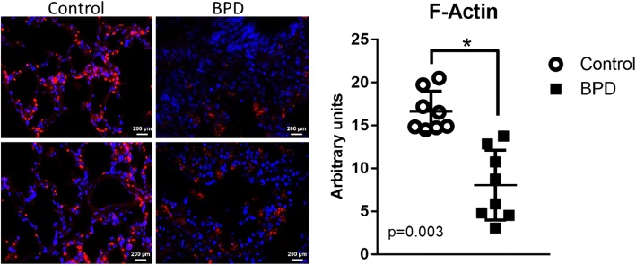
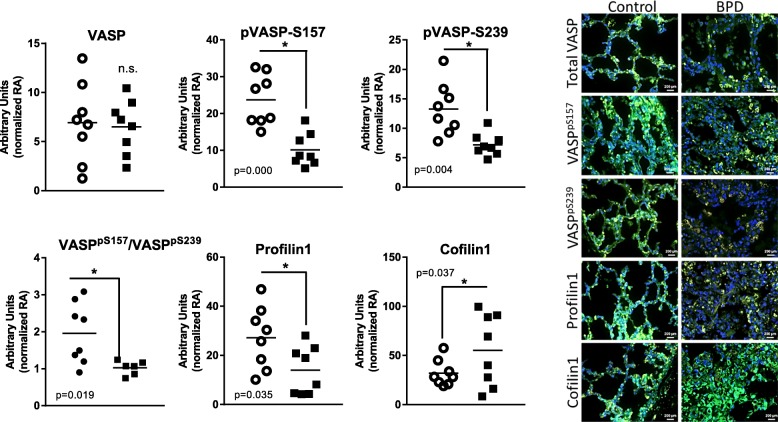
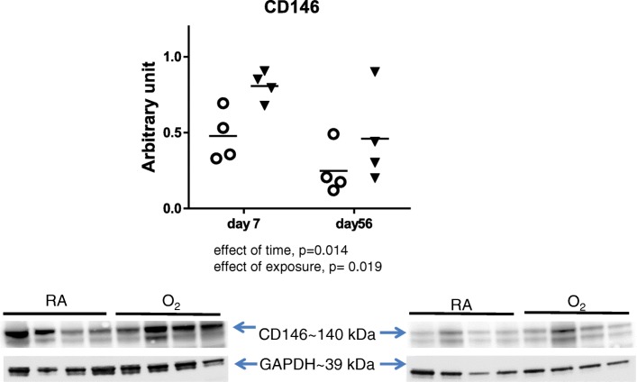
Results: Decreased F-actin content, ratio of VASPpS157/VASPpS239, and profilin 1 expression were observed in human lung tissues but this same pattern was not observed in lungs from hyperoxia-exposed newborn mice. Increases in cofilin1 expression were observed in both human and mouse tissues at 7d indicating a dysregulation in actin dynamics which may be related to altered growth. CD146 levels were elevated in human and newborn mice tissues (7d).

Conclusion: Altered phosphorylation of VASP and expression of profilin 1 and cofilin 1 in human tissues indicate that the pathophysiology of BPD involves dysregulation of actin binding proteins. Lack of similar changes in a mouse model of hyperoxia exposure imply that disruption in actin binding protein expression may be linked to interventions or morbidities other than hyperoxia alone.

Keywords: Actin binding proteins; BPD; CD146; Cofilin1; Hyperoxia; L-MSCs; Profilin1; VASP; VASPpS157; VASPpS239.


Conflict of interest statement

Ethics approval and consent to participate

Animal study protocols (#AR07–00028) were approved by the IACUC at The Research Institute at Nationwide Children’s Hospital. Human lung autopsy tissues from infants that died with BPD and infants that died at similar postnatal ages from non-pulmonary causes (control) were obtained from Dr. Gloria Pryhuber and collected as part of the BRINDL repository and Lung Map consortium.

Consent for publication

Not Applicable

Competing interests

The authors declare that they have no competing interests.

Publisher’s Note

Springer Nature remains neutral with regard to jurisdictional claims in published maps and institutional affiliations.

Figures

Fig. 1
Fig. 1
Human lung autopsy tissues F-actin content. Lung tissues sections were obtained from infants that died with BPD (closed squares) or infants that died at similar ages from non-pulmonary illnesses (open circles). Phalloidin used to stain F-actin in red while nuclei stained with DAPI in blue. Confocal microscopy images at 40X magnification processed in NIH Image-J software to measure F-actin contents. Four independent images were obtained from each slide and averaged for each individual creating n = 8 for each group. Statistical analysis was performed using Welch’s unequal variances t-test, and significance is indicated as *, p < 0.05; compared to controls
Fig. 2
Fig. 2
VASP phosphorylation, profilin1 and cofilin1 expression in human lung autopsy tissue. Lung tissues sections were obtained from infants that died with BPD (closed squares) or infants that died at similar ages from non-pulmonary illnesses (open circles). Fluorescent microscopy followed by NIH Image J analysis was used to quantify VASP expression and differential phosphorylation, and profilin1 and cofilin1 expression in human autopsy samples. Images are 40X magnification. Green, VASP, phosphor-VASP, profilin1 and cofilin1 stained with specific antibody against each protein; red, F-actin stained with phalloidin; and blue, nuclei stained with DAPI. Four independent images were obtained from each slide and averaged for each individual creating n = 8 for each group. Statistical analysis was performed using Welch’s unequal variances t-test, n = 8 in each group, and significance is indicated as *, p < 0.05; and n.s. = non significant compared to respective controls
Fig. 3
Fig. 3
VASP phosphorylation, profilin1 and cofilin1 level in lung tissue homogenates from mouse pups exposed to hyperoxia until day 7. Western blot analysis was performed on whole lung tissue homogenates obtained from mice exposed to room air (RA, open circles) or 85% O2 (O2, closed triangles) from birth through day 7 or at day 56 after 14 days of hyperoxia exposure and 42 days of room air recovery. Band intensity was quantified by densitometry and statistical analysis was performed using two-way ANOVA with Tukey’s post hoc, n = 4 in each group. *, p < 0.05 and n.s. = non significant
Fig. 4
Fig. 4
L-MSCs (CD146) levels in lung tissue homogenates from mouse pups exposed to hyperoxia. Western blot analysis was performed on whole lung tissue homogenates obtained from mice exposed to room air (RA, open circles) or 85% O2 (O2, closed triangles) at day 7 and 56. Band intensity was quantified by densitometry and statistical analysis was performed using two = way ANOVA with Tukey’s post-hoc. *, p < 0.05 and n.s. = non significant
Fig. 5
Fig. 5
L-MSCs marker (CD146) expression in human lung autopsy tissues. Lung tissues sections were obtained from infants that died with BPD (closed squares) or infants that died at similar ages from non-pulmonary illnesses (open circles). Green, CD146 stained with specific antibody; and blue, nuclei stained with DAPI. Confocal microscopy images at 40X magnification processed in NIH Image-J software. Statistical analysis was performed using Welch’s unequal variances t-test, n = 8 in each group, and significance is indicated as ***, p < 0.0001; compared to controls
Fig. 6
Fig. 6
Schematic representation of hypothesis. Hyperoxia exposure decreases VASP expression and phosphorylation disrupting actin dynamics in human tissues. Early responses include increases in cofilin1 in attempts to initiate repair. In later stages of injury and dysregulated repair, we observe decreases in VASP phosphorylation and profilin1 which are indicative of impaired repair. L-MSCs play a significant role in repair and proliferate in response to injury stimuli. Early increase in CD146 is indicative of enhanced L-MSCs proliferation and homing in mouse lungs, while at later stages decreased CD146 in human autopsy tissue is suggestive injury beyond repair. We speculated that disruptions in ABP dynamics may be due to interventions or morbidities other than hyperoxia alone in humans with BPD

Similar articles

Cited by

References

    1. Benz PM, Blume C, Seifert S, Wilhelm S, Waschke J, Schuh K, Gertler F, Munzel T, Renne T. Differential VASP phosphorylation controls remodeling of the actin cytoskeleton. J Cell Sci. 2009;122:3954–3965. doi: 10.1242/jcs.044537. - DOI - PMC - PubMed
    1. Aszodi A, Pfeifer A, Ahmad M, Glauner M, Zhou XH, Ny L, Andersson KE, Kehrel B, Offermanns S, Fassler R. The vasodilator-stimulated phosphoprotein (VASP) is involved in cGMP- and cAMP-mediated inhibition of agonist-induced platelet aggregation, but is dispensable for smooth muscle function. EMBO J. 1999;18:37–48. doi: 10.1093/emboj/18.1.37. - DOI - PMC - PubMed
    1. Doppler H, Storz P. Regulation of VASP by phosphorylation: consequences for cell migration. Cell Adhes Migr. 2013;7:482–486. doi: 10.4161/cam.27351. - DOI - PMC - PubMed
    1. Pula G, Krause M. Role of Ena/VASP proteins in homeostasis and disease. Handb Exp Pharmacol. 2008:39–65. - PubMed
    1. Siton O, Bernheim-Groswasser A. Reconstitution of actin-based motility by vasodilator-stimulated phosphoprotein (VASP) depends on the recruitment of F-actin seeds from the solution produced by cofilin. J Biol Chem. 2014;289:31274–31286. doi: 10.1074/jbc.M114.586958. - DOI - PMC - PubMed

MeSH terms

LinkOut - more resources