. 2018 Nov 21;37(1):279.
doi: 10.1186/s13046-018-0950-9.

LncRNA XIST/miR-34a axis modulates the cell proliferation and tumor growth of thyroid cancer through MET-PI3K-AKT signaling

Affiliations

LncRNA XIST/miR-34a axis modulates the cell proliferation and tumor growth of thyroid cancer through MET-PI3K-AKT signaling

Hua Liu et al. J Exp Clin Cancer Res. .

Abstract

Background: Thyroid cancer is one of the most prevalent malignancies in endocrine system. Further understanding and revealing the molecular mechanism underlying thyroid cancer are indispensable for the development of effective diagnosis and treatments. In the present study, we attempted to provide novel basis for targeted therapy for thyroid cancer from the aspect of lncRNA-miRNA-mRNA interaction.

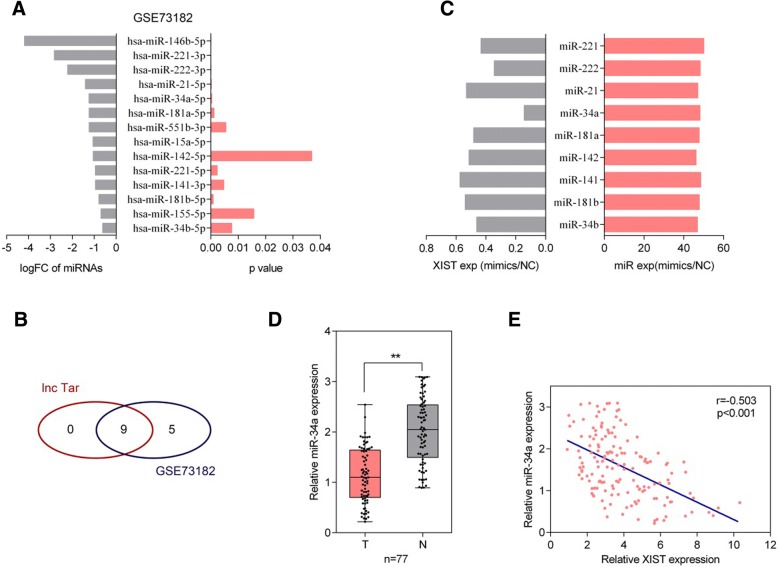
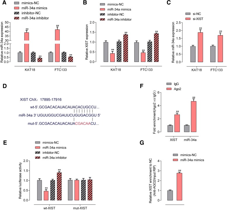
Methods: The expression and cellular function of XIST (X-inactive specific transcript) was determined. miRNAs which may be direct targets of XIST were screened for from online GEO database and miR-34a was selected. Next, the predicted binding between XIST and miR-34a, and the dynamic effect of XIST and miR-34a on downstream MET (hepatocyte growth factor receptor)-PI3K (phosphoinositide 3-kinase)-AKT (α-serine/threonine-protein kinase) signaling was evaluated.

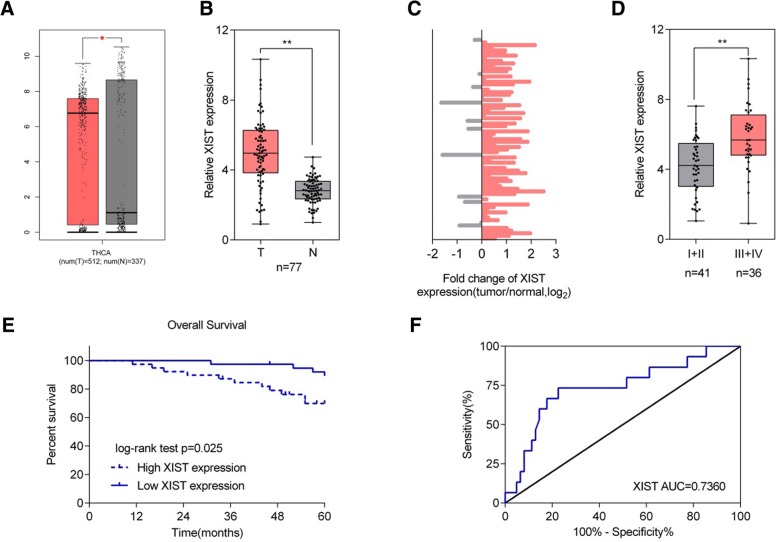
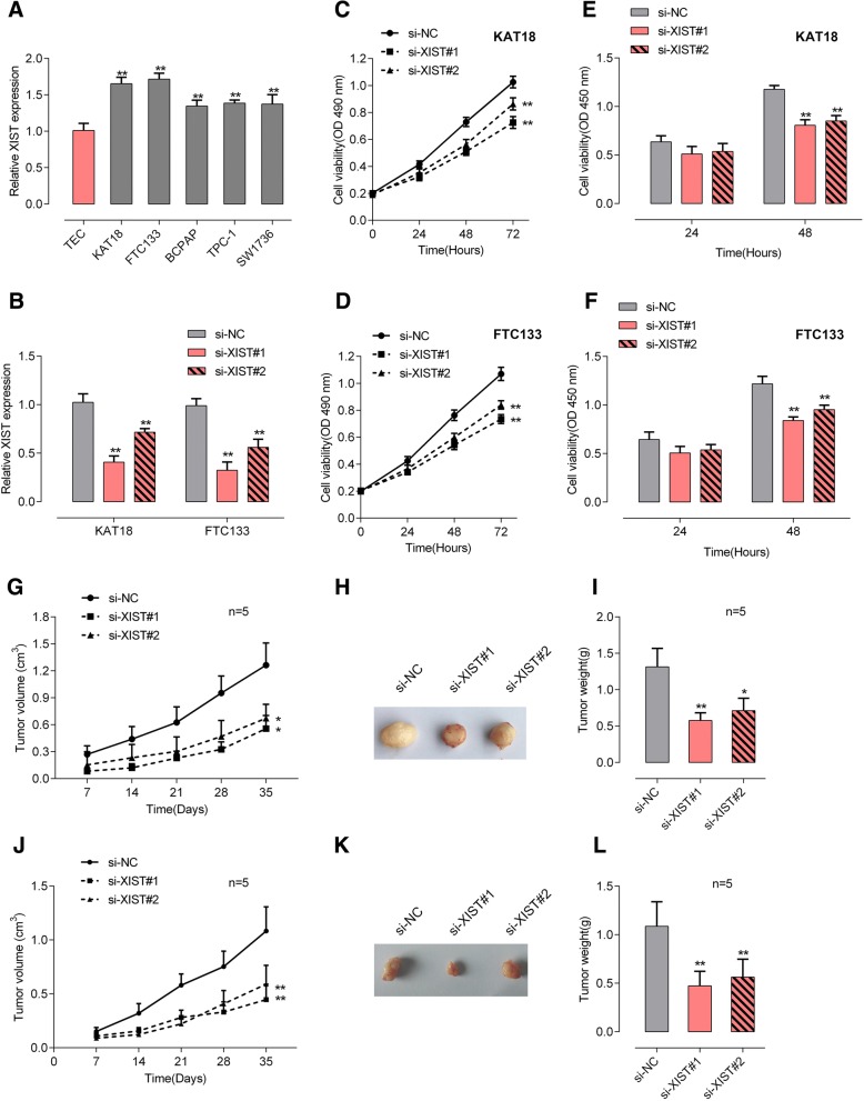
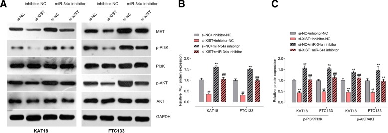
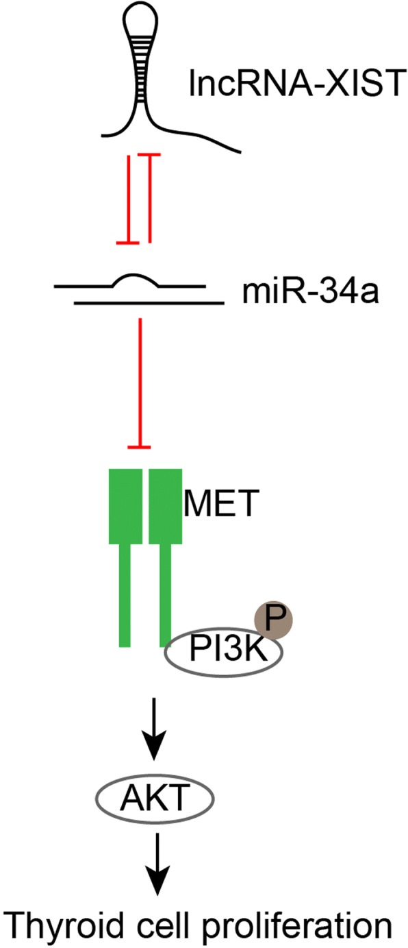
Results: XIST was significantly up-regulated in thyroid cancer tissues and cell lines; XIST knockdown suppressed the cell proliferation in vivo and the tumor growth in vitro. Based on online database and online tool prediction results, miR-34a was underexpressed in thyroid cancer and might be a direct target of XIST. Herein, we confirmed the negative interaction between XIST and miR-34a; moreover, XIST knockdown could reduce the protein levels of MET, a downstream target of miR-34a, and the phosphorylation of PI3K and AKT. In thyroid cancer tissues, MET mRNA and protein levels of MET were up-regulated; MET was positively correlated with XIST while negatively correlated with miR-34a, further confirming that XIST serves as a ceRNA for miR-34a through sponging miR-34a, competing with MET for miR-34a binding, and finally modulating thyroid cancer cell proliferation and tumor growth.

Conclusion: In the present study, we provided novel experimental basis for targeted therapy for thyroid cancer from the aspect of lncRNA-miRNA-mRNA interaction.

Keywords: MET; PI3K/AKT; Thyroid cancer; XIST; miR-34a.


Conflict of interest statement

Ethics approval and consent to participate

The study was conducted in accordance with the Declaration of Helsinki, and the protocol was approved by the Ethic Committee of Xiangya Hospital, Central South University. All of the enrolled lung cancer patients signed informed consent forms.

Consent for publication

Not applicable.

Competing interests

The authors declare that they have no competing interests.

Publisher’s Note

Springer Nature remains neutral with regard to jurisdictional claims in published maps and institutional affiliations.

Figures

Fig. 1
Fig. 1
Expression of XIST and its correlation with the clinical parameters in patients with thyroid cancer (a) XIST expression in 512 thyroid cancer tissue samples and 337 adjacent non-tumor tissue samples based on the data from TCGA-THCA were exhibited. b XIST expression in 77 paired thyroid cancer tissue samples and adjacent non-tumor tissue samples was examined using real-time PCR assays. c The distribution of XIST expression fold-change shown as tumor/normal (log2). d XIST expression in tumor tissues grouping by TNM stages (I + II vs. III + IV). The data are presented as mean ± SD of three independent experiments. *P < 0.05, **P < 0.01. e Kaplan-Meier overall survival curves for 77 patients with thyroid cancer classified according to relative XIST expression level. (F) Receiver operating characteristic (ROC) curve showing the area under the curve (AUC) to analyze the sensitivity and specificity of XIST as a biomarker for thyroid cancer
Fig. 2
Fig. 2
XIST knockdown inhibits the cell and tumor growth of thyroid cancer (a) XIST expression in five thyroid cancer cell lines (FTC133, BCPAP, TPC-1, SW1736 and KAT18) and a normal human thyroid epithelium cell line (TEC) was examined using real-time PCR assays. b XIST knockdown was achieved by transfection of si-XIST#1 or si-XIST#2, as confirmed using real-time PCR assays. c-d The cell viability was determined using MTT assays. e-f The DNA synthesis ability was determined using BrdU assays. g-i The tumor volume and tumor weight derived from si-XIST#1 or si-XIST#2-transfected KAT18 cells were examined. j-l The tumor volume and tumor weight derived from si-XIST#1 or si-XIST#2-transfected TPC-1 cells were examined. The data are presented as mean ± SD of three independent experiments. *P < 0.05, **P < 0.01
Fig. 3
Fig. 3
Screening and verification of candidate XIST downstream target miRNAs (a) The fold-change of down-regulated miRNAs in thyroid cancer compared to non-tumor tissue samples, showing as logFC and P values. b Candidate downstream miRNAs of XIST predicted by lnc Tar and the most down-regulated miRNAs reported by GSE73182. (c) The expression of the above candidate miRNAs was achieved by transfection of miRNA mimics, as confirmed using real-time PCR assays; XIST expression was then determined using real-time PCR assays. d miR-34a expression in 77 paired thyroid cancer and non-tumor tissue samples was examined using real-time PCR assays. The data are presented as mean ± SD of three independent experiments. **P < 0.01. e The correlation between miR-34a expression and XIST expression was analyzed using Spearman’s rank correlation analysis
Fig. 4
Fig. 4
XIST targets miR-34a to negatively interact with miR-34a (a) KAT18 and FTC133 cells were transfected with miR-34a mimics or miR-34a inhibitor to achieve miR-34a expression, as confirmed using real-time PCR assays. b XIST expression was determined using real-time PCR assays. c miR-34a expression in response to XIST knockdown was determined using real-time PCR assays. d-e Luciferase reporter gene assays were performed to confirm the direct targeting of XIST to miR-34a. f Association of miR-34a and XIST with AGO2 in HEK293 cells. Detection of AGO2 and IgG using Immunoblotting assays. g RIP assay in HEK293 cells transfected with control miRNA (miR-NC) or miR-34a followed by real-time PCR to detect XIST associated with AGO2. The data are presented as mean ± SD of three independent experiments. **P < 0.01
Fig. 5
Fig. 5
XIST modulates miR-34a downstream MET-PI3K-AKT signaling via miR-34a (a-c) KAT18 and FTC113 cells were co-transfected with si-XIST and miR-34a inhibitor; the protein levels of MET, p-PI3K, PI3K, p-AKT and AKT were examined using Immunoblotting assays. The data are presented as mean ± SD of three independent experiments. **P < 0.01, compared to control group; ##P < 0.01, compared to si-XIST group
Fig. 6
Fig. 6
MET expression in tissue samples and its correlation with XIST and miR-34a (a) MET mRNA expression in 512 thyroid cancer and 337 non-tumor tissues based on TCGA-THCA database. b MET mRNA expression in 77 paired thyroid cancer and non-tumor tissue samples was determined using real-time PCR assays. The data are presented as mean ± SD of three independent experiments. *P < 0.05, **P < 0.01. c The protein levels of MET in three paired randomly selected thyroid cancer and non-tumor tissues were determined using Immunoblotting assays. d-e The correlation between XIST and MET, between miR-34a and MET was analyzed using Spearman’s rank correlation analysis
Fig. 7
Fig. 7
Mechanism map

Similar articles

Cited by

References

    1. Zaballos MA, Santisteban P. Key signaling pathways in thyroid cancer. J Endocrinol. 2017;235(2):R43–R61. doi: 10.1530/JOE-17-0266. - DOI - PubMed
    1. Colin IM, Denef JF, Lengele B, Many MC, Gerard AC. Recent insights into the cell biology of thyroid angiofollicular units. Endocr Rev. 2013;34(2):209–238. doi: 10.1210/er.2012-1015. - DOI - PMC - PubMed
    1. Fusco A, Grieco M, Santoro M, Berlingieri MT, Pilotti S, Pierotti MA, Della Porta G, Vecchio G. A new oncogene in human thyroid papillary carcinomas and their lymph-nodal metastases. Nature. 1987;328(6126):170–172. doi: 10.1038/328170a0. - DOI - PubMed
    1. Nikiforova MN, Chiosea SI, Nikiforov YE. MicroRNA expression profiles in thyroid tumors. Endocr Pathol. 2009;20(2):85–91. doi: 10.1007/s12022-009-9069-z. - DOI - PubMed
    1. Zhang X, Mao H, Lv Z. MicroRNA role in thyroid cancer pathogenesis. Front Biosci (Landmark Ed) 2013;18:734–739. doi: 10.2741/4135. - DOI - PubMed

MeSH terms