. 2018 Nov 21;15(1):323.
doi: 10.1186/s12974-018-1354-7.

STING-mediated type-I interferons contribute to the neuroinflammatory process and detrimental effects following traumatic brain injury

Affiliations

STING-mediated type-I interferons contribute to the neuroinflammatory process and detrimental effects following traumatic brain injury

Amar Abdullah et al. J Neuroinflammation. .

Abstract

Background: Traumatic brain injury (TBI) represents a major cause of disability and death worldwide with sustained neuroinflammation and autophagy dysfunction contributing to the cellular damage. Stimulator of interferon genes (STING)-induced type-I interferon (IFN) signalling is known to be essential in mounting the innate immune response against infections and cell injury in the periphery, but its role in the CNS remains unclear. We previously identified the type-I IFN pathway as a key mediator of neuroinflammation and neuronal cell death in TBI. However, the modulation of the type-I IFN and neuroinflammatory responses by STING and its contribution to autophagy and neuronal cell death after TBI has not been explored.

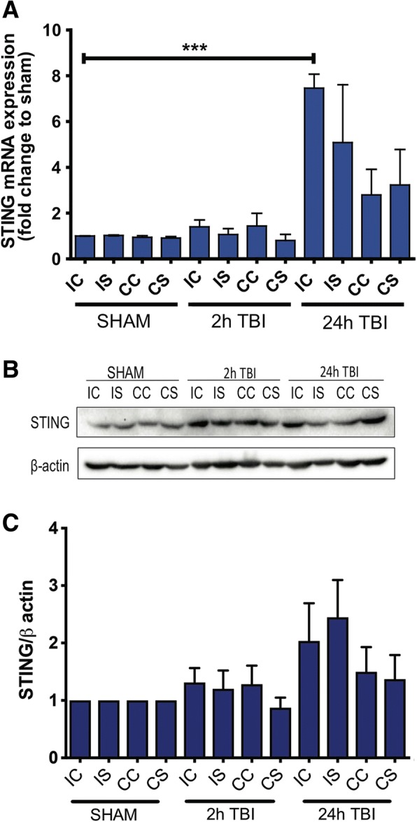
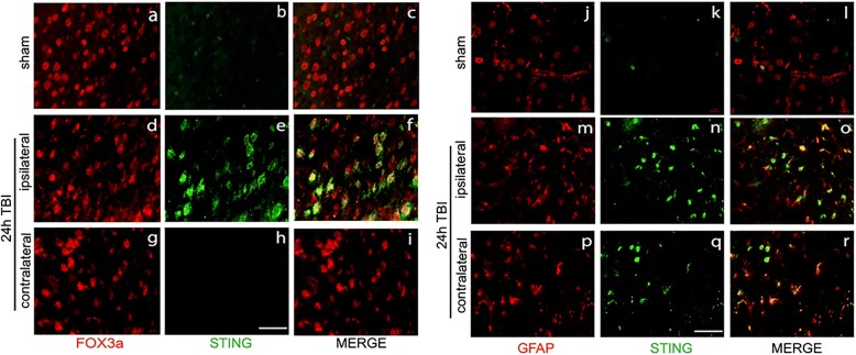
Methods: C57BL/6J wild-type (WT) and STING-/- mice (8-10-week-old males) were subjected to controlled cortical impact (CCI) surgery and brains analysed by QPCR, Western blot and immunohistochemical analyses at 2 h or 24 h. STING expression was also analysed by QPCR in post-mortem human brain samples.

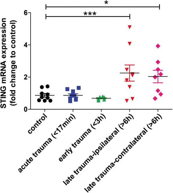
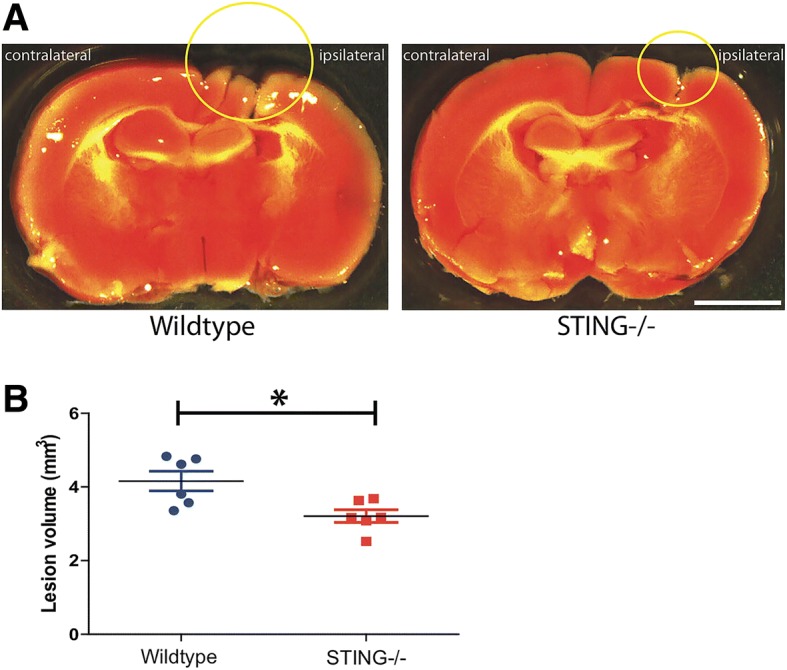
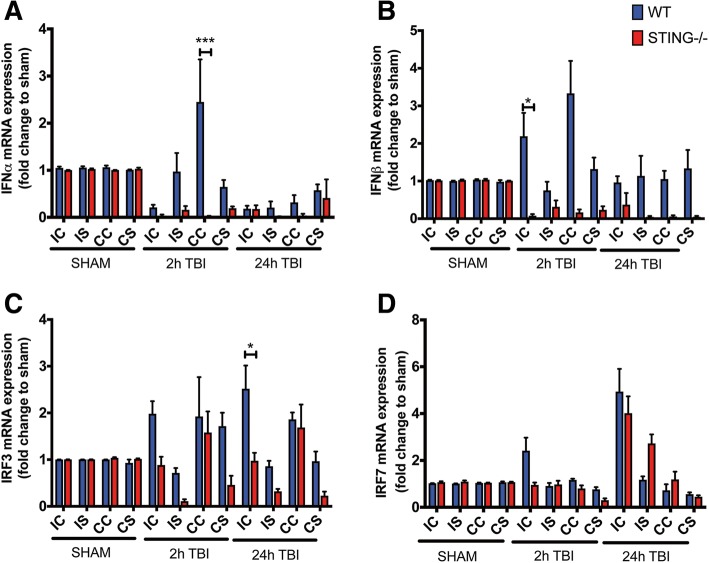
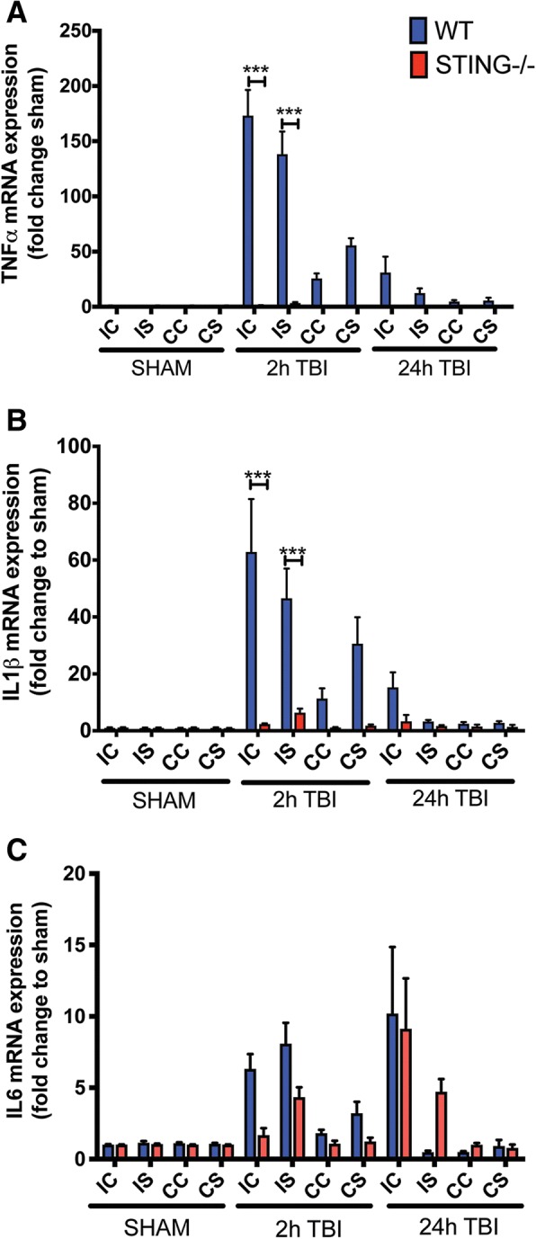
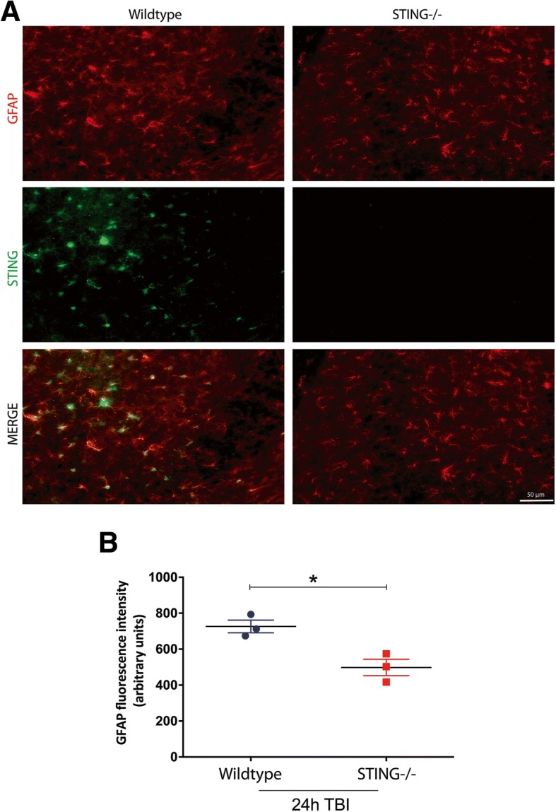
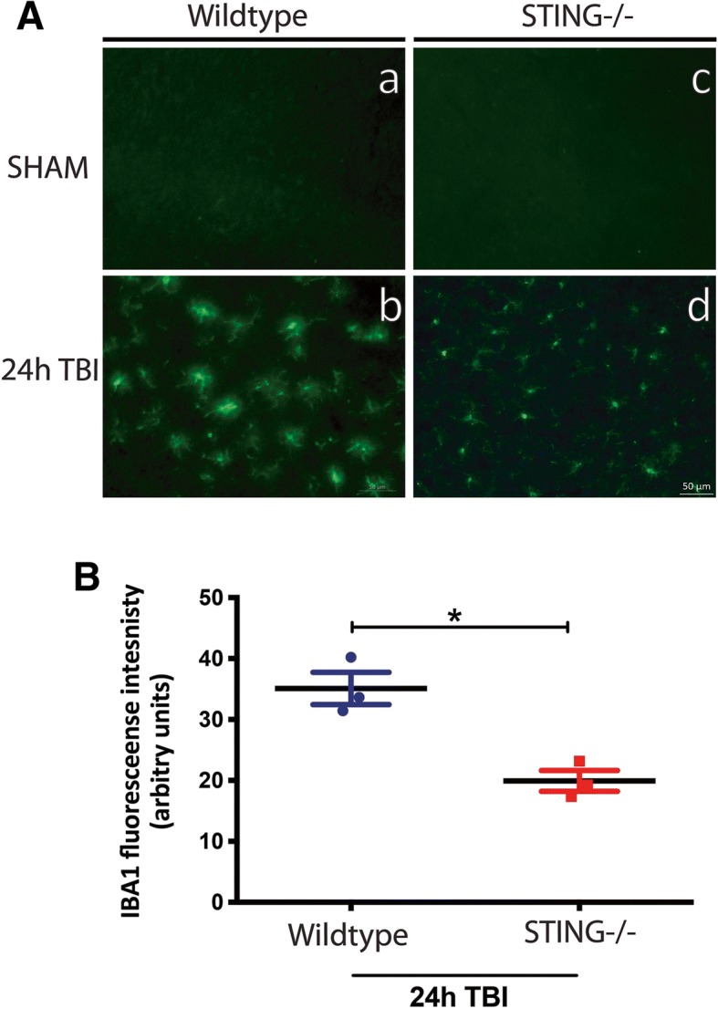
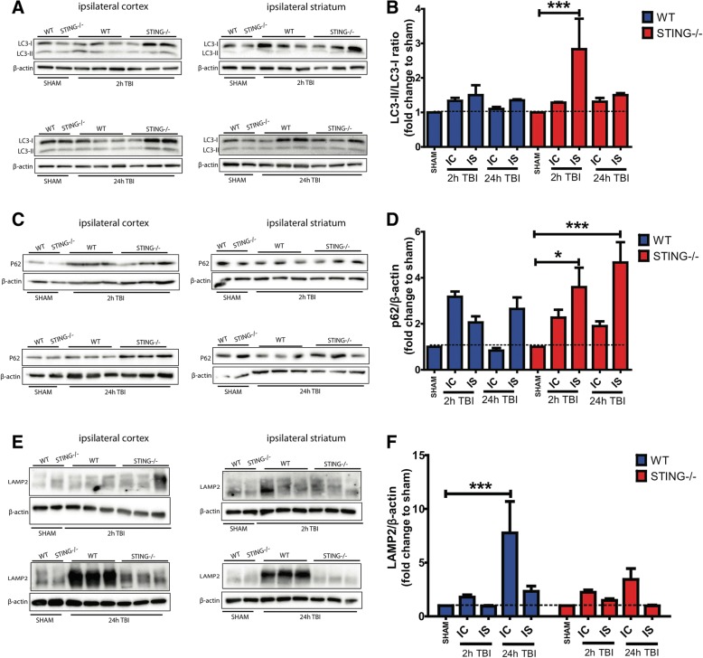
Results: A significant upregulation in STING expression was identified in late trauma human brain samples that was confirmed in wild-type mice at 2 h and 24 h after CCI. This correlated with an elevated pro-inflammatory cytokine profile with increased TNF-α, IL-6, IL-1β and type-I IFN (IFN-α and IFN-β) levels. This expression was suppressed in the STING-/- mice with a smaller lesion volume in the knockout animals at 24 h post CCI. Wild-type mice also displayed increased levels of autophagy markers, LC3-II, p62 and LAMP2 after TBI; however, STING-/- mice showed reduced LAMP2 expression suggesting a role for STING in driving dysfunctional autophagy after TBI.

Conclusion: Our data implicates a detrimental role for STING in mediating the TBI-induced neuroinflammatory response and autophagy dysfunction, potentially identifying a new therapeutic target for reducing cellular damage in TBI.

Keywords: Autophagy; Neuroinflammation; STING; Traumatic brain injury; Type-I interferon.


Conflict of interest statement

Ethics approval and consent to participate

All experiments involving animal surgery were conducted in accordance with University of Melbourne Animal Ethics Committee (#1212477.1). All procedures with human tissues were conducted in accordance with the Australian National Health & Medical Research Council’s National Statement on Ethical Conduct in Human Research (2007), the Victorian Human Tissue Act 1982, the National Code of Ethical Autopsy Practice and the Victorian Government Policies and Practices in Relation to Postmortem.

Consent for publication

Not applicable

Competing interests

The authors declare that they have no competing interests.

Publisher’s Note

Springer Nature remains neutral with regard to jurisdictional claims in published maps and institutional affiliations.

Figures

Fig. 1
Fig. 1
STING mRNA expression is upregulated in post-mortem human trauma brain samples. QPCR analysis identified increased mRNA expression of STING in late trauma group of post-mortem human trauma brains compared with controls (n = 8–10). Data represents mean ± SEM, *p < 0.05; ***p < 0.0001
Fig. 2
Fig. 2
TBI induces STING expression in WT mice. Increased STING mRNA was detected by QPCR as shown in a (n = 6 for each time point) and Western blot analysis b (n = 3). Quantification of STING protein expression in (b) shown in (c). All data is expressed as mean ± SEM, ***p < 0.001. IC ipsilateral cortex, IS ipsilateral striatum, CC contralateral cortex, CS contralateral striatum
Fig. 3
Fig. 3
TBI induces STING expression in both neurons and astrocytes. Representative images of immunohistochemical analysis showing brain sections from the wild-type sham (ac and jl) and CCI (di and mr) mice co-labelled with antibodies to identify neurons (FOX3a-positive cells; ai) and astrocyte (GFAP-positive cell; jr). Images are taken near lesion area (n = 3). Scale bar = 100 μM
Fig. 4
Fig. 4
Genetic ablation of STING confers neuroprotection 24 h after TBI. Total lesion volumes of wild-type and STING−/− mice were assessed by TTC staining and quantified using Image J. a Representative images demonstrating reduced infarct size in STING−/− compared to wild-type mice 24 h after CCI (n = 6). b Quantification of (a) showing STING−/− mice have significantly reduced lesion volumes compared to WT mice 24 h after CCI. Data represents mean ± SEM, p < *0.05; n = 6 animals per group. Scale bar: 1 mm
Fig. 5
Fig. 5
STING−/− mice display reduced type-I IFN signalling after TBI. QPCR analysis identified increased mRNA expression of IRF3 (a), IRF7 (b), IFNα (c) and IFN-β (d) in WT brains mice as compared to STING−/− mice after CCI (n = 6). Data represents mean ± SEM, *p < 0.05; ***p < 0.0001. IC ipsilateral cortex, IS ipsilateral striatum, CC contralateral cortex, CS contralateral striatum
Fig. 6
Fig. 6
STING−/− mice exhibit reduced TBI-induced pro-inflammatory cytokines in vivo. mRNA expression of IL-1β (a), IL-6 (b) and TNF-α (c) in brain tissue of wild-type and STING−/− mice subjected to CCI. (n = 6 mice for each timepoint). Data represents mean ± SEM, *p < 0.05; **p < 0.01; ***p < 0.001. IC ipsilateral cortex, IS ipsilateral striatum, CC contralateral cortex, CS contralateral striatum
Fig. 7
Fig. 7
STING−/− mice display reduced GFAP protein expression following TBI. a Representative images (n = 6 mice for each genotype and timepoint) showing GFAP protein levels in wild-type and STING−/− mice after TBI as assessed by western blot analysis. Significantly increased expression of GFAP was observed in the ipsilateral striatum of wild-type mice 24 h post-TBI but not in STING−/− mice as compared with sham group, as quantified in (b). Data represent mean ± SEM, *p < 0.05. IC ipsilateral cortex, IS ipsilateral striatum, CC contralateral cortex, CS contralateral striatum
Fig. 8
Fig. 8
STING−/− mice exhibit reduced GFAP immunostaining compared with WT mice after TBI. a High-power GFAP (red) staining co-labelled with STING (green) in the ipsilateral side of the brain. GFAP intensity is measured in (b) showing reduced GFAP immuno-reactivity in the STING−/− mice 24 h after TBI (n = 3 mice for each genotype and timepoint). Data represent mean ± SEM, *p < 0.05
Fig. 9
Fig. 9
STING−/− brains exhibit ramified microglial morphologies and reduced IBA-1 immunostaining following TBI. 24 h after TBI, brains from STING−/− mice displayed microglia with ramified morphologies as identified by IBA-1 staining (d) as compared to WT mice (b). IBA-1 expression was significantly reduced in the STING−/− mice at 24 h post-TBI as quantified in (b) (n = 3 mice for each genotype and timepoint)
Fig. 10
Fig. 10
STING−/− brains exhibit ramified microglial flux 24 h post-TBI. a LC3, (c) p62 and (e) LAMP2 expression was detected in wild-type and STING−/− brains (n = 6) by Western blot. LC3-I, LC3-II, p62 and LAMP2 levels were normalised to β-actin levels respectively. For densitometry calculations, b LC3-II/LC3-I ratio, d p62/β-actin ratio and f LAMP2/β-actin ratio was then determined from these values and was calculated as a fold change relative to genotype sham control. Data is expressed as mean ± SEM, **p < 0.01; ***p < 0.001. IC ipsilateral cortex, IS ipsilateral striatum

References

    1. Lingsma HF, Roozenbeek B, Steyerberg EW, Murray GD, Maas AIR. Early prognosis in traumatic brain injury: from prophecies to predictions. Lancet Neurol. 2010;9:543–554. doi: 10.1016/S1474-4422(10)70065-X. - DOI - PubMed
    1. Kim DY, O'Leary M, Nguyen A, Kaji A, Bricker S, Neville A, Bongard F, Putnam B, Plurad D. The effect of platelet and desmopressin administration on early radiographic progression of traumatic intracranial hemorrhage. J Neurotrauma. 2015;32:1815–1821. doi: 10.1089/neu.2014.3728. - DOI - PubMed
    1. Skolnick BE, Maas AI, Narayan RK, van der Hoop RG, MacAllister T, Ward JD, Nelson NR, Stocchetti N. A clinical trial of progesterone for severe traumatic brain injury. N Engl J Med. 2014;371:2467–2476. doi: 10.1056/NEJMoa1411090. - DOI - PubMed
    1. Wright DW, Yeatts SD, Silbergleit R, Palesch YY, Hertzberg VS, Frankel M, Goldstein FC, Caveney AF, Howlett-Smith H, Bengelink EM, et al. Very early administration of progesterone for acute traumatic brain injury. N Engl J Med. 2014;371:2457–2466. doi: 10.1056/NEJMoa1404304. - DOI - PMC - PubMed
    1. Blennow K, Hardy J, Zetterberg H. The neuropathology and neurobiology of traumatic brain injury. Neuron. 2012;76:886–899. doi: 10.1016/j.neuron.2012.11.021. - DOI - PubMed

MeSH terms