. 2018 Nov 21;9(1):326.
doi: 10.1186/s13287-018-1052-5.

Human UCB-MSCs treatment upon intraventricular hemorrhage contributes to attenuate hippocampal neuron loss and circuit damage through BDNF-CREB signaling

Affiliations

Human UCB-MSCs treatment upon intraventricular hemorrhage contributes to attenuate hippocampal neuron loss and circuit damage through BDNF-CREB signaling

Hyo Rim Ko et al. Stem Cell Res Ther. .

Abstract

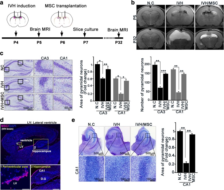
Background: Human umbilical cord blood-derived mesenchymal stem cells (hUCB-MSCs) have been shown to prevent brain damage and improve neurocognition following intraventricular hemorrhage (IVH). However, the molecular mechanisms underlying the effects of hUCB-MSCs are still elusive. Thus, as the hippocampus is essential for learning, memory, and cognitive functions and is intimately involved in the ventricular system, making it a potential site of IVH-induced injury, we determined the molecular basis of the effects of hUCB-derived MSCs on hippocampal neurogenesis and the recovery of hippocampal neural circuits after IVH in a rodent model.

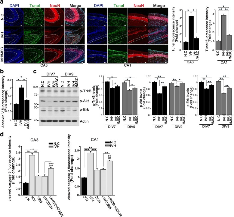
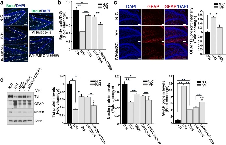
Methods: We inflicted severe IVH injury on postnatal day 4 (P4) in rats. After confirmation of successful induction of IVH using MRI (P5), intracerebroventricular administration of MSCs (ICV-MSC) was performed at 2 days post-injury (P6). For hippocampal synaptic determination, a rat entorhinal-hippocampus (EH) organotypic slice co-culture (OSC) was performed using day 3 post-IVH brains (P7) with or without ICV-MSCs. A similar strategy of experiments was applied to those rats receiving hUCB-MSC transfected with BDNF-Si-RNA for knockdown of BDNF or scrambled siRNA controls after IVH. The molecular mechanism of the MSCs effects on neurogenesis and the attenuation of neuron death was determined by evaluation of BDNF-TrkB-Akt-CREB signaling axis.

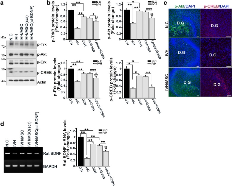
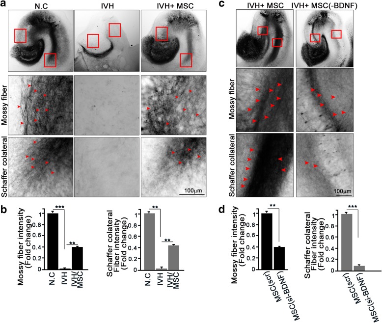
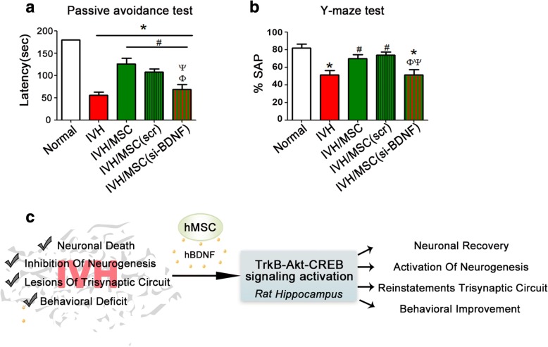
Results: We showed that treatment with hUCB-MSCs attenuated neuronal loss and promoted neurogenesis in the hippocampus, an area highly vulnerable to IVH-induced brain injury. hUCB-MSCs activate BDNF-TrkB receptor signaling, eliciting intracellular activation of Akt and/or Erk and subsequent phosphorylation of CREB, which is responsible for promoting rat BDNF transcription. In addition to the beneficial effects of neuroprotection and neurogenesis, hUCB-MSCs also contribute to the restoration of impaired synaptic circuits in the hippocampus and improve neurocognitive functions in IVH-injured neonatal rat through BDNF-TrkB-CREB signaling axis activation.

Conclusions: Our data suggest that hUCB-MSCs possess therapeutic potential for treating neuronal loss and neurocognitive dysfunction in IVH through the activation of intracellular TrkB-CREB signaling that is invoked by hUCB-MSC-secreted BDNF.

Keywords: BDNF; CREB; Hippocampus; Intraventricular hemorrhage; Mesenchymal stem cells.


Conflict of interest statement

Ethics approval

This study was reviewed and approved by the Institutional Animal Care and Use Committee (IACUC) of Sungkyunkwan University School of Medicine (SUSM) (code 17-6-4-1). SUSM is an Association for Assessment and Accreditation of Laboratory Animal Care International (AAALAC International; No. 001004)-accredited facility and abide by the Institute of Laboratory Animal Resources (ILAR) guide. All experimental procedures were carried out in accordance with the regulations of the IACUC guideline of Sungkyunkwan University.

Consent for publication

Not applicable.

Competing interests

Won Soon Park and Yun Sil Chang declare potential conflicts of interest arising from a filed or issued patent titled “Composition for treating intraventricular hemorrhage in preterm infants comprising mesenchymal stem cells” as co-inventors, not as patentees.

Publisher’s Note

Springer Nature remains neutral with regard to jurisdictional claims in published maps and institutional affiliations.

Figures

Fig. 1
Fig. 1
hUCB-MSC treatment prevents neuron loss in the hippocampus after severe IVH injury. a, b In P4 rat pups, IVH was induced by IC injection of a total of 200 μl of fresh maternal whole blood (100 μl each into the right and left ventricles). Normal control rats received a sham operation without IC blood injection. At P5 (1 day after IVH), severe intraventricular hemorrhage (IVH) was confirmed by brain magnetic resonance imaging (MRI), and IVH rat pups showing minimal or non-visible IVH on the brain MRI were excluded. At P32 (26 days after MSC treatment), IVH damage and its recovery by MSC treatment were measured by MRI. c At P7 after animal modeling of intraventricular hemorrhaging, rat brain was embedded in paraffin. Serial hippocampal coronal sections were stained with cresyl violet (left). The area of pyramidal neurons and number of pyramidal neurons were quantified using ImageJ software (right). d After severe intraventricular hemorrhage and transplantation of hUCB-MSCs, P7 rat brain was fixed with 4% PFA. Mesenchymal stem cells were stained with anti-human mitochondria antibody. Scale bar, 50 μm (periventricular zone), 500 μm (hippocampus). D.G: dentate gyrus. e In P7 rats, entorhinal-hippocampal slice cultures were prepared after intraventricular hemorrhage. Slices are stained with cresyl violet at DIV7 (left). The area of pyramidal neurons was quantified in a black box in ImageJ software (right). MSC markedly protected against neuronal cell death in the CA1 region of the IVH model. Images shown here are representative of at least three independent experiments (n ≥ 15 per group). Data are shown as mean ± SEM. *p < 0.05, **p < 0.005 versus the indicated group
Fig. 2
Fig. 2
hUCB-MSC treatment promotes neuronal survival in the hippocampus through activation of BDNF signaling. a TUNEL staining and immunohistochemistry were performed using sections of paraffin block (P7). DNA was stained with DAPI (blue). TUNEL-positive nuclear staining is green. Neurons were stained with NeuN, a neuronal marker (red). Representative images show the hippocampal CA1 and CA3 regions (left). Scale bar, 50 μm. Quantification of relative immunofluorescence intensity of TUNEL signal is shown as a bar graph (right). Data are shown as mean ± SEM; *p < 0.05 and ** p < 0.005 versus the indicated group (n ≥ 15 per group). b Rat brain paraffin sections were stained with annexin V and NeuN. Bar graph shows the annexin V fluorescence intensity. A representative figure is shown in Additional file 1: Figure S1A. Images shown here are representative of at least three independent experiments. Data are shown as mean ± SEM. *p < 0.05 versus the indicated group. c Whole rat brains were dissected and lysed at the indicated time points and applied for immunoblotting analysis with the indicated antibodies. Bar graph shows quantified amounts of p-TrkB, pAKT, and p-Erk. Data are representative of at least three independent experiments. Data are shown as mean ± SEM; *p < 0.05 and **p < 0.005 versus the indicated group. d Paraffin sections were stained with cleaved caspase3 and NeuN. Bar graph shows the cleaved caspase 3 fluorescence intensity. Data are shown as mean ± SEM from three independent experiments, and a representative image is shown in Additional file 1: Figure S1D. n ≥ 15 per group. **p < 0.005 and ***p < 0.0005 versus the indicated group. ###p < 0.0005 versus N.C, ΦΦΦp < 0.0005 versus IVH
Fig. 3
Fig. 3
hUCB-MSC treatment enhanced neurogenesis in the hippocampus after neuron loss. a, b Effect of MSCs on neurogenesis in the hippocampal dentate gyrus following IVH. At 3 days after severe intraventricular hemorrhage, rat was injected intraperitoneally with 5-bromo-deoxyuridine (BrdU, 100 mg/kg). At 24 h after BrdU i.p. injection, the rat brain was fixed with 4% PFA for 48 h. To determine neurogenesis, a paraffin section was stained with anti-BrdU antibody (green) and DAPI (blue). Scale bar, 100 μm. Data are shown as mean ± SEM; **p < 0.005 versus the indicated group. b Quantification of BrdU+ signal is shown as a bar graph. Images shown here are representative of at least three independent experiments, and each value represents the mean ± SEM of triplicate measurements. *p < 0.05 and **p < 0.005 versus the indicated group. c After severe IVH and transplantation of hUCB-MSCs, P7 rat brain was fixed with 4% PFA for 48 h. Glial cells were stained with anti-GFAP antibody (red). Nuclei were counterstained with DAPI (blue). Scale bar, 100 μm. The bar graph shows GFAP fluorescence intensity. Data are shown as mean ± SEM; n = 3. *p < 0.05 versus the indicated group (right). d Rat hippocampi of IVH/MSC rat models were dissected at p7. Lysates were subjected to IB with the indicated antibodies. Data are representative of three independent experiments. Data are shown as mean ± SEM; n ≥ 15 per group. *p < 0.05 and **p < 0.005 versus the indicated group. ##p < 0.005 versus N.C, ΦΦp < 0.005 versus IVH
Fig. 4
Fig. 4
The BDNF-Akt/Erk-CREB axis contributes to neurogenesis in the hippocampus after IVH injury. a, b Rat hippocampus were dissected and lysed at p7 in IVH/MSC rat models. Lysates were subjected to IB with the indicated antibodies (left). Amounts of p-TrkB, p-Akt, p-Erk, and p-CREB protein were determined by IB (right). Images shown here are representative of at least three independent experiments, and each value represents the mean ± SEM of triplicate measurements; *p < 0.05 and ** p < 0.005 versus indicated groups, ##p < 0.005 versus N.C, ###p < 0.0005 versus N.C, Φp < 0.05 versus IVH, ΦΦp < 0.005 versus IVH. c Hippocampal slice cultures were prepared after IVH. Slices were cultured for an additional 7 days and fixed with 4% paraformaldehyde. Slice was stained with p-ATK and p-CREB antibody (green). Nuclei were counterstained with DAPI stain (blue). Scale bar, 50 μm. d Rat hippocampus was dissected at P7. RNA was isolated from the hippocampus, and the mRNA levels of rat BDNF were determined using species-specific primers (left). Quantification of rat BDNF mRNA measurements from three independent experiments is shown on the right. Data are shown as mean ± SEM; **p < 0.005 versus the indicated group. ##p < 0.005 versus N.C, ΦΦp < 0.005 versus IVH
Fig. 5
Fig. 5
hUCB-MSC treatment contributes to the recovery of IVH injury-mediated lesions in the hippocampal trisynaptic circuit. a, b Hippocampal slices of IVH/MSC rat models were cultured at P7. The anterograde axonal tracer biocytin was placed on the entorhinal cortex, D.G, and CA3 at DIV8 and fixed with 4% PFA. Biocytin was visualized using the ABC-DAB method. Red arrows indicate mossy fibers (upper) and Schaffer collateral fibers (bottom). Image shown here is representative of at least three independent experiments. Scale bar, 100 μm. b Bar graph shows mossy fiber intensity and Schaffer collateral fiber intensity. Data are shown as mean ± SEM; n ≥ 20 per group. **p < 0.005 and ***p < 0.0005 versus the indicated group. c To determine the effect of MSCs (BDNF) on hippocampal trisynaptic fiber, MSCs (scr) or MSCs (si-BDNF) were transplanted at p6 following IVH. After 7 days of slice culture, the anterograde axonal tracer biocytin was placed on the slice. Red arrows indicate the mossy (middle) and Schaffer collateral (bottom) fibers. Scale bar, 100 μm. d Bar graph shows mossy fiber intensity and Schaffer collateral fiber intensity. Image shown here is representative of at least three independent experiments. Data are shown as mean ± SEM; **p < 0.005 and ***p < 0.0005 versus the indicated group
Fig. 6
Fig. 6
hUCB-MSC treatment reinstates synaptic function through BDNF signaling. Learning and memory functional outcomes on passive avoidance test (a) and Y-maze (b) at P32. Data are expressed as mean ± standard error of the mean (SEM). *p < 0.05 versus normal, #p < 0.05 versus IVH control, Φp < 0.05 versus IVH+MSC, Ψp < 0.05 versus IVH+scrambled siRNA-transfected MSCs. c Schematic diagram of hUCB-MSC-induced BDNF signaling after IVH in rat hippocampus

References

    1. Payne AH, Hintz SR, Hibbs AM, Walsh MC, Vohr BR, Bann CM, Wilson-Costello DE, Eunice Kennedy Shriver National Institute of Child Health. Human Development Neonatal Research Network Neurodevelopmental outcomes of extremely low-gestational-age neonates with low-grade periventricular-intraventricular hemorrhage. JAMA Pediatr. 2013;167:451–459. doi: 10.1001/jamapediatrics.2013.866. - DOI - PMC - PubMed
    1. Ballabh P. Intraventricular hemorrhage in premature infants: mechanism of disease. Pediatr Res. 2010;67:1–8. doi: 10.1203/PDR.0b013e3181c1b176. - DOI - PMC - PubMed
    1. Philip AG, Allan WC, Tito AM, Wheeler LR. Intraventricular hemorrhage in preterm infants: declining incidence in the 1980s. Pediatrics. 1989;84:797–801. - PubMed
    1. Jain RK. Molecular regulation of vessel maturation. Nat Med. 2003;9:685–693. doi: 10.1038/nm0603-685. - DOI - PubMed
    1. Wilson-Costello D, Friedman H, Minich N, Fanaroff AA, Hack M. Improved survival rates with increased neurodevelopmental disability for extremely low birth weight infants in the 1990s. Pediatrics. 2005;115:997–1003. doi: 10.1542/peds.2004-0221. - DOI - PubMed

Publication types

MeSH terms