. 2018 Nov 21;9(1):319.
doi: 10.1186/s13287-018-1068-x.

Stem cell library screen identified ruxolitinib as regulator of osteoblastic differentiation of human skeletal stem cells

Affiliations

Stem cell library screen identified ruxolitinib as regulator of osteoblastic differentiation of human skeletal stem cells

Nihal AlMuraikhi et al. Stem Cell Res Ther. .

Abstract

Background: Better understanding of the signaling pathways that regulate human bone marrow stromal stem cell (hBMSC) differentiation into bone-forming osteoblasts is crucial for their clinical use in regenerative medicine. Chemical biology approaches using small molecules targeting specific signaling pathways are increasingly employed to manipulate stem cell differentiation fate.

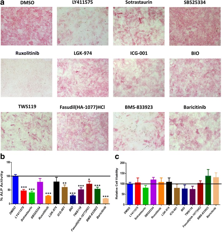
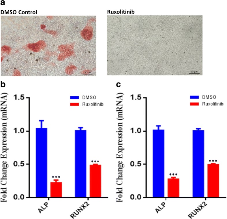
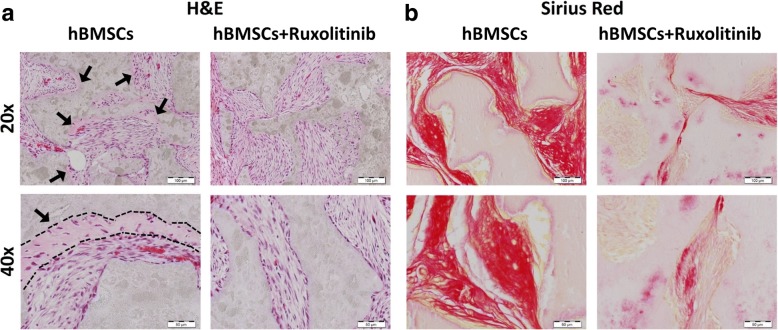
Methods: We employed alkaline phosphatase activity and staining assays to assess osteoblast differentiation and Alizarin R staining to assess mineralized matrix formation of cultured hBMSCs. Changes in gene expression were assessed using an Agilent microarray platform, and data normalization and bioinformatics were performed using GeneSpring software. For in vivo ectopic bone formation experiments, hMSCs were mixed with hydroxyapatite-tricalcium phosphate granules and implanted subcutaneously into the dorsal surface of 8-week-old female nude mice. Hematoxylin and eosin staining and Sirius Red staining were used to detect bone formation in vivo.

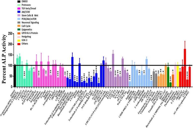
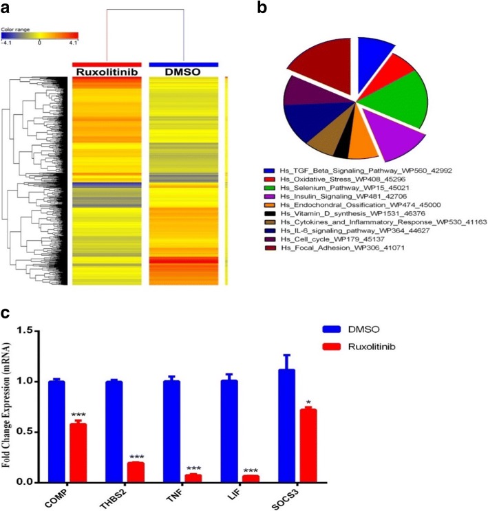
Results: We identified several compounds which inhibited osteoblastic differentiation of hMSCs. In particular, we identified ruxolitinib (INCB018424) (3 μM), an inhibitor of JAK-STAT signaling that inhibited osteoblastic differentiation and matrix mineralization of hMSCs in vitro and reduced ectopic bone formation in vivo. Global gene expression profiling of ruxolitinib-treated cells identified 847 upregulated and 822 downregulated mRNA transcripts, compared to vehicle-treated control cells. Bioinformatic analysis revealed differential regulation of multiple genetic pathways, including TGFβ and insulin signaling, endochondral ossification, and focal adhesion.

Conclusions: We identified ruxolitinib as an important regulator of osteoblast differentiation of hMSCs. It is plausible that inhibition of osteoblast differentiation by ruxolitinib may represent a novel therapeutic strategy for the treatment of pathological conditions caused by accelerated osteoblast differentiation and mineralization.


Conflict of interest statement

Ethics approval and consent to participate

All animal experiments received the appropriate ethical approval from the King Saud University Ethical Research Committee.

Consent for publication

Not applicable.

Competing interests

The authors declare that they have no competing interests.

Publisher’s Note

Springer Nature remains neutral with regard to jurisdictional claims in published maps and institutional affiliations.

Figures

Fig. 1
Fig. 1
Functional screen of stem cell signaling small molecule library for their effects on osteoblast differentiation of human bone marrow stromal stem cells (hBMSCs). hBMSCs were induced into osteoblasts for 10 days in the presence of the indicated small molecule inhibitors (3.0 μM) or DMSO vehicle control. Data are presented as mean alkaline phosphatase (ALP) activity ± SEM, n ≥ 10 from three independent experiments. Small molecules are grouped according to their targeted signaling pathway. DMSO dimethyl sulfoxide. *P < 0.05; **P < 0.05; ***P < 0.0005
Fig. 2
Fig. 2
The effect of a selected panel of small molecules targeting multiple signaling pathways on osteoblast differentiation of hBMSCs. a Representative alkaline phosphatase (ALP) staining of hBMSCs on day 10 following treatment with the indicated compounds (concentration 3.0 μM). Images were taken at × 10 magnification using a Zeiss inverted microscope. b Quantification of ALP activity in hBMSCs following treatment with the indicated compounds (concentration 3 μM) versus vehicle-treated control cells at day 10. Data are presented as mean percentage ALP activity ± SEM, n > 16. **P < 0.05; ***P < 0.0005. c Cell viability assay using alamarBlue showing the relative cell viability in hBMSCs following treatment with the indicated compounds (3 μM) versus vehicle-treated control cells on day 10 post-osteoblast differentiation. Abbreviations: ALP alkaline phosphatase, DMSO dimethyl sulfoxide
Fig. 3
Fig. 3
The effect of ruxolitinib on osteoblastic differentiation of hBMSCs. a hMSCs were induced into osteoblasts for 21 days in the absence (left panel) or presence (right panel) of ruxolitinib and were stained for mineralized matrix formation using Alizarin Red stain. Images were taken at × 10 magnification using a Zeiss inverted microscope. Quantitative RT-PCR analysis for gene expression of alkaline phosphatase (ALP) and RUNX2 in hBMSCs inducted into osteoblasts for 10 days (b) or 21 days (c) in the absence (blue) or presence (red) of ruxolitinib. Cells treated with DMSO were used as control. Gene expression was normalized to β-actin. Data are presented as mean fold change ± SEM (n = 6) from two independent experiments. ***P ≤ 0.0005. Abbreviations: ALP alkaline phosphatase, RUNX2 runt-related transcription factor 2, DMSO dimethyl sulfoxide
Fig. 4
Fig. 4
Ruxolitinib affects multiple pathways during osteoblastic differentiation of hBMSCs. a Heat map analysis and unsupervised hierarchical clustering performed on differentially expressed genes during osteoblast differentiation of ruxolitinib-treated compared to DMSO-treated control hBMSCs. b Pie chart illustrating the distribution of selected enriched pathway categories for the downregulated genes identified in osteoblast differentiated ruxolitinib-treated hBMSCs compared to DMSO-treated control cells. c Validation of a selected panel of downregulated genes during osteoblastic differentiation of ruxolitinib-treated hBMSCs compared to DMSO-treated control cells using qRT-PCR. Gene expression was normalized to β-actin. Data are presented as mean fold change ± SEM (n = 6) from two independent experiments; *P < 0.05; ***P < 0.0005
Fig. 5
Fig. 5
Ruxolitinib inhibits in vivo ectopic bone formation. Ruxolitinib-treated and control hBMSCs were implanted with hydroxyl apatite/tricalcium phosphate (HA/TCP) subcutaneously into NOD/SCID mice. The histology of in vivo bone formation was examined with H&E (a) and Sirius red (b) staining. Black arrows indicate the bone formation (× 20), and black line shows the bone formed zone with osteoblast between the HA and spindle-shaped hMSCs (× 40). Images were taken at × 20 (first row; scale bar = 100 μm) and × 40 (second row; scale bar = 50 μm) magnification using a light microscope. Abbreviation: H&E hematoxylin and eosin

References

    1. Aldahmash A, Zaher W, Al-Nbaheen M, Kassem M. Human stromal (mesenchymal) stem cells: basic biology and current clinical use for tissue regeneration. Ann Saudi Med. 2012;32(1):68–77. doi: 10.5144/0256-4947.2012.68. - DOI - PMC - PubMed
    1. Elsafadi M, Manikandan M, Almalki S, Mobarak M, Atteya M, Iqbal Z, Hashmi JA, Shaheen S, Alajez N, Alfayez M, et al. TGFbeta1-induced differentiation of human bone marrow-derived MSCs is mediated by changes to the actin cytoskeleton. Stem Cells Int. 2018;2018:6913594. doi: 10.1155/2018/6913594. - DOI - PMC - PubMed
    1. Qiu W, Andersen TE, Bollerslev J, Mandrup S, Abdallah BM, Kassem M. Patients with high bone mass phenotype exhibit enhanced osteoblast differentiation and inhibition of adipogenesis of human mesenchymal stem cells. J Bone Miner Res. 2007;22(11):1720–1731. doi: 10.1359/jbmr.070721. - DOI - PubMed
    1. Jafari A, Siersbaek MS, Chen L, Qanie D, Zaher W, Abdallah BM, Kassem M. Pharmacological inhibition of protein kinase G1 enhances bone formation by human skeletal stem cells through activation of RhoA-Akt signaling. Stem Cells. 2015;33(7):2219–2231. doi: 10.1002/stem.2013. - DOI - PubMed
    1. Fakhry M, Hamade E, Badran B, Buchet R, Magne D. Molecular mechanisms of mesenchymal stem cell differentiation towards osteoblasts. World J Stem Cells. 2013;5(4):136–148. doi: 10.4252/wjsc.v5.i4.136. - DOI - PMC - PubMed

Publication types