. 2018 Nov 21;20(1):259.
doi: 10.1186/s13075-018-1754-1.

Aberrant expression of interleukin-23-regulated miRNAs in T cells from patients with ankylosing spondylitis

Affiliations

Aberrant expression of interleukin-23-regulated miRNAs in T cells from patients with ankylosing spondylitis

Ning-Sheng Lai et al. Arthritis Res Ther. .

Abstract

Background: Interleukin (IL)-23 can facilitate the differentiation of IL-17-producing helper T cells (Th17). The IL-23/IL-17 axis is known to play a key role in the immunopathogenesis of ankylosing spondylitis (AS). We hypothesized that the expression of microRNAs (miRNAs, miRs) would be regulated by IL-23 and that these miRNAs could participate in the immunopathogenesis of AS.

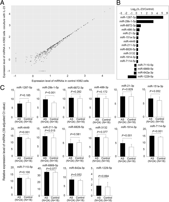
Methods: Expression profiles of human miRNAs in K562 cells, cultured in the presence or absence of IL-23 for 3 days, were analyzed by microarray. Potentially aberrantly expressed miRNAs were validated using T-cell samples from 24 patients with AS and 16 control subjects. Next-generation sequencing (NGS) was conducted to search for gene expression and biological functions regulated by specific miRNAs in the IL-23-mediated signaling pathway.

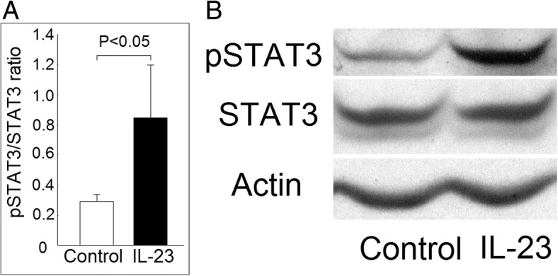
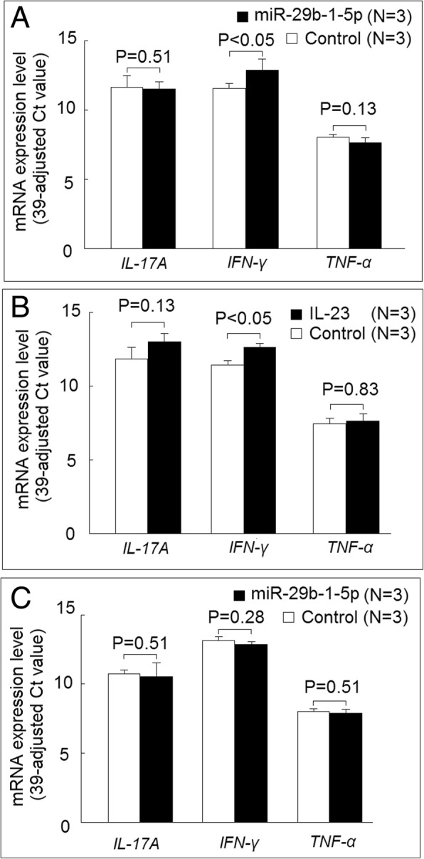
Results: Initial analysis revealed that the expression levels of 12 miRNAs were significantly higher, whereas those of 4 miRNAs were significantly lower, in K562 cells after coculture with IL-23 for 3 days. Among these IL-23-regulated miRNAs, the expression levels of miR-29b-1-5p, miR-4449, miR-211-3p, miR-1914-3p, and miR-7114-5p were found to be higher in AS T cells. The transfection of miR-29b-1-5p mimic suppressed IL-23-mediated signal transducer and activator of transcription 3 (STAT3) phosphorylation in K562 cells. After NGS analysis and validation, we found that miR-29b-1-5p upregulated the expression of angiogenin, which was also upregulated in K562 cells after coculture with IL-23. Increased expression of miR-29b-1-5p or miR-211-3p could enhance interferon-γ expression.

Conclusions: Among the miRNAs regulated by IL-23, expression levels of five miRNAs were increased in T cells from patients with AS. The transfection of miR-29b-1-5p mimic could inhibit the IL-23-mediated STAT3 phosphorylation and might play a role in negative feedback control in the immunopathogenesis of AS.

Keywords: Angiogenin; Ankylosing spondylitis; IL-23; MicroRNAs; STAT3; T cells.


Conflict of interest statement

Ethics approval and consent to participate

This study was conducted in accordance with the Declaration of Helsinki and was approved by the institutional review board of Buddhist Dalin Tzu Chi Hospital, Taiwan (no. B10502002). Signed informed consent was obtained from all patients.

Consent for publication

Not applicable.

Competing interests

The authors declare that they have no competing interests.

Publisher’s Note

Springer Nature remains neutral with regard to jurisdictional claims in published maps and institutional affiliations.

Figures

Fig. 1
Fig. 1
Effect of interleukin (IL)-23 on signal transducer and activator of transcription 3 (STAT3) protein phosphorylation in K562 cells. a The phosphorylation ratio of STAT3 increased in K562 cells after coculture with IL-23 (20 ng/ml) for 3 days compared with those cocultured with medium (control) only. b A representative case
Fig. 2
Fig. 2
Altered expression of interleukin (IL)-23-regulated microRNAs (miRNAs) in T cells from patients with ankylosing spondylitis (AS) and from healthy control subjects. a Expression profiles of miRNAs in K562 cells cocultured with or without IL-23 (20 ng/ml) for 3 days, evaluated using microarray analysis. Each scatter spot represents the mean raw signals of miRNA in three repeats of each treatment. b The expression levels of 12 miRNAs were significantly higher, whereas the expression levels of 4 miRNAs were significantly lower, in K562 cells after coculture with IL-23 for 3 days (P < 0.05). c Increased expression of miR-29b-1-5p, miR-4449, miR-211-3p, miR-1914-3p, and miR-7114-5p in AS T-cell miRNA, compared with normal T cells after validation
Fig. 3
Fig. 3
Effect of miR-29b-1-5p on signal transducer and activator of transcription 3 (STAT3) protein phosphorylation in K562 cells. a Increased miR-29b-1-5p expression in K562 cells after transfection with miR-29b-1-5p mimic versus scramble oligonucleotides. b The phosphorylation ratio of STAT3 decreased in K562 cells after transfection with miR-29b-1-5p mimic compared with those transfected with scramble oligonucleotides after culturing with medium for 48 h. c A representative case
Fig. 4
Fig. 4
Identification of miR-29b-1-5p-regulated genes. a Expression profiles of messenger RNAs (mRNAs) in K562 cells transfected with miR-29b-1-5p mimic or scramble oligonucleotides then cultured with medium for 48 h were evaluated using RNA-Seq transcriptome analysis. Each scatter spot represents the mean raw signals of microRNA (miRNA) in three repeats of each treatment. b The RNA expression levels of 16 genes were significantly higher, whereas the RNA expression levels of 10 genes were significantly lower, in K562 cells after being transfected with miR-29b-1-5p mimic for 48 h. c Four genes potentially involved in the inflammatory responses were selected for further analysis. The mRNA expression levels of ANG were significantly elevated in K562 cells after coculture with interleukin (IL)-23 (20 ng/ml) for 3 days (Fig. 3c)
Fig. 5
Fig. 5
Protein expression of angiogenin was regulated by miR-29b-1-5p and interleukin (IL)-23. a The protein expression of angiogenin was elevated in K562 cells after being transfected with miR-29b-1-5p mimic for 48 h compared with those transfected with scramble oligonucleotides. b A representative case. c The protein expression of angiogenin was also increased in K562 cells after coculture with IL-23 for 3 days compared with those cultured with medium only. d A representative case
Fig. 6
Fig. 6
Effect of miR-29b-1-5p on proinflammatory cytokine expression. a The messenger RNA (mRNA) expression levels of interferon-γ (IFN-γ), but not interleukin (IL)-17A or tumor necrosis factor-α (TNF-α), were increased in K562 cells 24 h after transfection of miR-29b-1-5p mimic compared with those transfected with scrambled oligonucleotides. b The mRNA expression levels of IFN-γ, but not IL-17A or TNF-α, were increased in K562 cells after coculture with IL-23 (20 ng/ml) compared with those cultured with medium alone. c The K562 cells transfected with miR-29b-1-5p mimic or scrambled oligonucleotides were cultured with IL-23 (20 ng/ml) for 24 h, and there was no difference in the mRNA expression levels of IFN-γ, IL-17A, or TNF-α between these two groups
Fig. 7
Fig. 7
Effect of miR-211-3p in proinflammatory cytokine expression. a The expression levels of miR-211-3p dramatically increased after transfection with miR-211-3p mimic compared with those transfected with scrambled oligonucleotides (control). b There were no differences in the mRNA expression levels of interferon-γ (IFN-γ), interleukin (IL)-17A, or tumor necrosis factor-α (TNF-α) in K562 cells 24 h after transfection of miR-211-3p mimic compared with those transfected with scrambled oligonucleotides. c The K562 cells transfected with miR-211-3p mimic or scrambled oligonucleotides were cultured with IL-23 (20 ng/ml) for 24 h. The mRNA expression levels of IFN-γ, but not IL-17A or TNF-α, were increased in those transfected with miR-211-3p mimic compared with the control

Similar articles

Cited by

References

    1. Bakland G, Gran JT, Nossent JC. Increased mortality in ankylosing spondylitis is related to disease activity. Ann Rheum Dis. 2011;70:1921–1925. doi: 10.1136/ard.2011.151191. - DOI - PubMed
    1. Sieper J, Poddubnyy D. Axial spondyloarthritis. Lancet. 2017;390:73–84. doi: 10.1016/S0140-6736(16)31591-4. - DOI - PubMed
    1. Ranganathan V, Gracey E, Brown MA, Inman RD, Haroon N. Pathogenesis of ankylosing spondylitis - recent advances and future directions. Nat Rev Rheumatol. 2017;13:359–367. doi: 10.1038/nrrheum.2017.56. - DOI - PubMed
    1. Brewerton DA, Hart FD, Nicholls A, Caffrey M, James DC, Sturrock RD. Ankylosing spondylitis and HL-A 27. Lancet. 1973;1:904–907. doi: 10.1016/S0140-6736(73)91360-3. - DOI - PubMed
    1. Wellcome Trust Case Control Consortium. Australo-Anglo-American Spondylitis Consortium (TASC) Burton PR, Clayton DG, Cardon LR, Craddock N, et al. Association scan of 14,500 nonsynonymous SNPs in four diseases identifies autoimmunity variants. Nat Genet. 2007;39:1329–1337. doi: 10.1038/ng.2007.17. - DOI - PMC - PubMed

Publication types