. 2018 Nov 21;9(1):317.
doi: 10.1186/s13287-018-1065-0.

Co-transplantation of mesenchymal stem cells improves spermatogonial stem cell transplantation efficiency in mice

Affiliations

Co-transplantation of mesenchymal stem cells improves spermatogonial stem cell transplantation efficiency in mice

Prashant Kadam et al. Stem Cell Res Ther. .

Abstract

Background: Spermatogonial stem cell transplantation (SSCT) could become a fertility restoration tool for childhood cancer survivors. However, since in mice, the colonization efficiency of transplanted spermatogonial stem cells (SSCs) is only 12%, the efficiency of the procedure needs to be improved before clinical implementation is possible. Co-transplantation of mesenchymal stem cells (MSCs) might increase colonization efficiency of SSCs by restoring the SSC niche after gonadotoxic treatment.

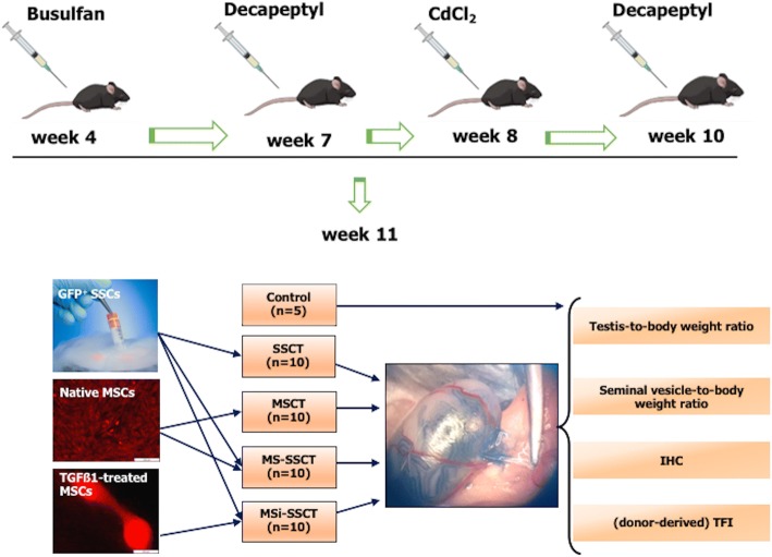
Methods: A mouse model for long-term infertility was developed and used to transplant SSCs (SSCT, n = 10), MSCs (MSCT, n = 10), a combination of SSCs and MSCs (MS-SSCT, n = 10), or a combination of SSCs and TGFß1-treated MSCs (MSi-SSCT, n = 10).

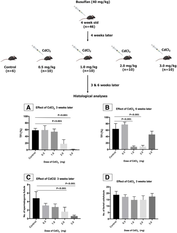
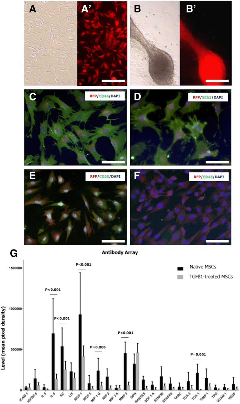
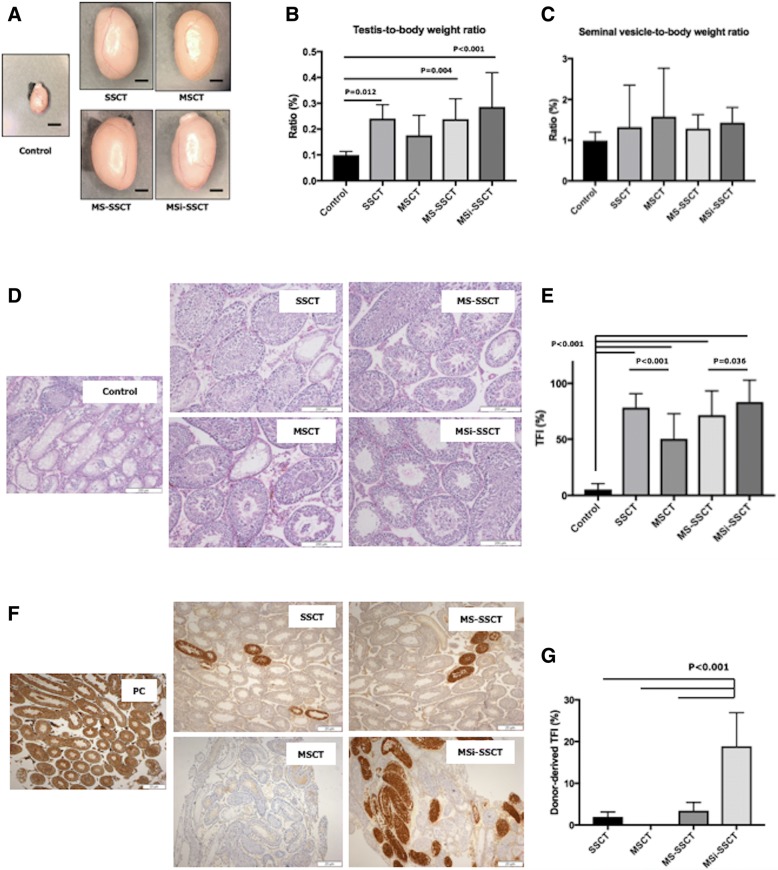
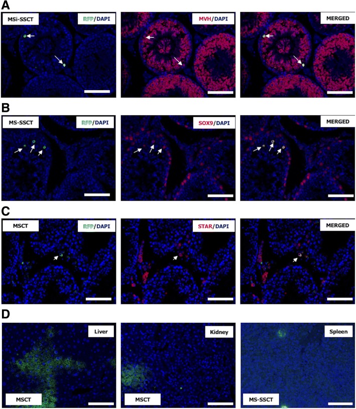
Results: The best model for transplantation was obtained after intraperitoneal injection of busulfan (40 mg/kg body weight) at 4 weeks followed by CdCl2 (2 mg/kg body weight) at 8 weeks of age and transplantation at 11 weeks of age. Three months after transplantation, spermatogenesis resumed with a significantly better tubular fertility index (TFI) in all transplanted groups compared to non-transplanted controls (P < 0.001). TFI after MSi-SSCT (83.3 ± 19.5%) was significantly higher compared to MS-SSCT (71.5 ± 21.7%, P = 0.036) but did not differ statistically compared to SSCT (78.2 ± 12.5%). In contrast, TFI after MSCT (50.2 ± 22.5%) was significantly lower compared to SSCT (P < 0.001). Interestingly, donor-derived TFI was found to be significantly improved after MSi-SSCT (18.8 ± 8.0%) compared to SSCT (1.9 ± 1.1%; P < 0.001), MSCT (0.0 ± 0.0%; P < 0.001), and MS-SSCT (3.4 ± 1.9%; P < 0.001). While analyses showed that both native and TGFß1-treated MSCs maintained characteristics of MSCs, the latter showed less migratory characteristics and was not detected in other organs.

Conclusion: Co-transplanting SSCs and TGFß1-treated MSCs significantly improves the recovery of endogenous SSCs and increases the homing efficiency of transplanted SSCs. This procedure could become an efficient method to treat infertility in a clinical setup, once the safety of the technique has been proven.

Keywords: Fertility restoration; Infertility; Mesenchymal stem cells; Spermatogonial stem cells; Transplantation.


Conflict of interest statement

Ethics approval and consent to participate

All procedures performed on the animals were in accordance with the ethical standards of the Federation for Laboratory Animal Science Associations guidelines and approved by the Institutional Animal Care and Use Committee of the Vrije Universiteit Brussel (VUB) Brussels, Belgium (14-216-2 and 16-216-3). Consent to participate is not applicable.

Consent for publication

Not applicable.

Competing interests

The authors declare that they have no competing interests.

Publisher’s Note

Springer Nature remains neutral with regard to jurisdictional claims in published maps and institutional affiliations.

Figures

Fig. 1
Fig. 1
Optimization of the mouse model for infertility. Several doses of CdCl2(0.5, 1.0, 2.0, and 3.0 mg/kg body weight) were tested in combination with busulfan (40 mg/kg body weight). TFI was assessed at two different time points after CdCl2 injection: (A) at week 3 and (B) week 6. (C) Graph showing quantitative analysis of spermatogonia per seminiferous tubule and (D) Sertoli cells per seminiferous tubule after CdCl2 treatment 3 weeks after injection
Fig. 2
Fig. 2
Mesenchymal stem cell culture. RFP+ MSCs were cultured in the absence (A, A′) or presence of TGFß1 (B, B′) for 15–21 days. MSCs showed a spindle-shaped morphology (A and B bright field in; A′ and B′ fluorescence microscope with Texas red filter). TGFß1-treated MSCs occasionally showed spheroid-like colonies. MSCs were positive for CD44 (C), SCA1 (D), and CD29 (E) and negative for CD45 (F). (G) Cytokine secretion profiles were obtained by incubating antibody array membranes with conditioned media from TGFß1-treated and non-treated MSC cultures (at 3 weeks). A, A′, B, B′ Scale bar = 200 μm. CF Scale bar = 100 μm
Fig. 3
Fig. 3
Transplantations. Transplantations were performed in GFP busulfan- and CdCl2-treated mice using SSCs from 5- to 7-day-old GFP+ mice and/or RFP+ MSCs. SSCT spermatogonial stem cell transplantation (n = 10), MSCT mesenchymal stem cell transplantation (n = 10), MS-SSCT transplantation of mesenchymal stem cells together with spermatogonial stem cells (n = 10), MSi-SSCT transplantation of TGFß1-treated mesenchymal stem cells together with spermatogonial stem cells (n = 10). Control (n = 5) mice received busulfan and CdCl2 but were not transplanted. Decapeptyl (4.26 mg/kg) was injected subcutaneously 1 week before CdCl2 injection and secondly 1 week before transplantations
Fig. 4
Fig. 4
Testicular histology 3 months after transplantation. The significant increase of testis size (a) and testis-to-body weight ratio (b) was found in transplanted groups compared to controls (control vs. SSCT, P = 0.012; control vs. MS-SSCT, P = 0.004, and control vs. MSi-SSCT, P < 0.001). However, no difference was observed for the seminal vesicle-to-body weight ratio (c). Resumption of spermatogenesis was found to be significantly improved in all transplanted groups compared to controls (P < 0.001) (d, e); Tubular fertility index (TFI) did not differ between SSCT and MSi-SSCT groups (e). Donor-derived spermatogenesis was confirmed with immunohistochemistry for GFP (f). Donor-derived TFI was found to be significantly different between SSCT and MSi-SSCT (P < 0.001), MSCT and MSi-SSCT (P < 0.001), and MS-SSCT and MSi-SSCT (P < 0.001) (g). SSCT, spermatogonial stem cell transplantation; MSCT, mesenchymal stem cell transplantation; MS-SSCT, transplantation of mesenchymal stem cells together with spermatogonial stem cells; MSi-SSCT, transplantation of TGFß1-treated mesenchymal stem cells together with spermatogonial stem cells. a Scale bar = 2 mm
Fig. 5
Fig. 5
Detection of transplanted mesenchymal stem cells. a Transplanted RFP+ MSCs (arrows) expressed a the germ cell marker MVH in all transplantation groups (MSCT, MS-SSCT, and MSi-SSCT). b The Sertoli cell marker SOX9 or c the Leydig cell marker STAR was expressed in transplanted MSCs after MSCT and MS-SSCT, but not after MSi-SSCT. d RFP+ MSCs (green) could also be detected in the liver, kidney, and spleen. Scale bar = 50 μm

Similar articles

Cited by

References

    1. Ward E, Desantis C, Robbins A, Kohler B, Jemal A. Childhood and adolescent cancer statistics, 2014. CA Cancer J Clin. 2014;64:83–103. doi: 10.3322/caac.21219. - DOI - PubMed
    1. Miller KD, Siegel RL, Lin CC, Mariotto AB, Kramer JL, Rowland JH, et al. Cancer treatment and survivorship statistics, 2016. CA Cancer J Clin. 2016;66:271–289. doi: 10.3322/caac.21349. - DOI - PubMed
    1. Wasilewski-Masker K, Seidel KD, Leisenring W, Mertens AC, Shnorhavorian M, Ritenour CW, et al. Male infertility in long-term survivors of pediatric cancer: a report from the childhood cancer survivor study. J Cancer Surviv. 2014;8:437–447. doi: 10.1007/s11764-014-0354-6. - DOI - PMC - PubMed
    1. Picton HM, Wyns C, Anderson RA, Goossens E, Jahnukainen K, Kliesch S, et al. A European perspective on testicular tissue cryopreservation for fertility preservation in prepubertal and adolescent boys. Hum Reprod. 2015;30:2463–2475. doi: 10.1093/humrep/dev190. - DOI - PubMed
    1. Goossens E, Tournaye H. Adult stem cells in the human testis. Semin Reprod Med. 2013;31:39–48. doi: 10.1055/s-0032-1331796. - DOI - PubMed

Publication types