

Thickness of the buccal bone wall and root angulation in the maxilla and mandible: an approach to cone beam computed tomography
- PMID: 30463614
- PMCID: PMC6249849
- DOI: 10.1186/s12903-018-0652-x
Thickness of the buccal bone wall and root angulation in the maxilla and mandible: an approach to cone beam computed tomography
Abstract
Background: The objective of this paper is to anatomically describe the bone morphology in the maxillary and mandibular tooth areas, which might help in planning post-extraction implants.
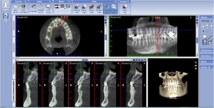
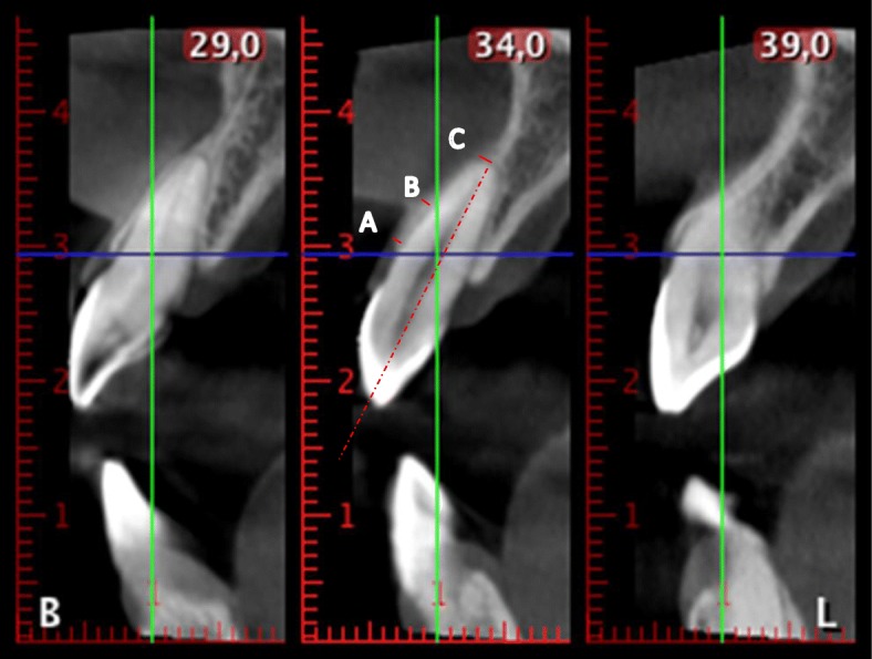
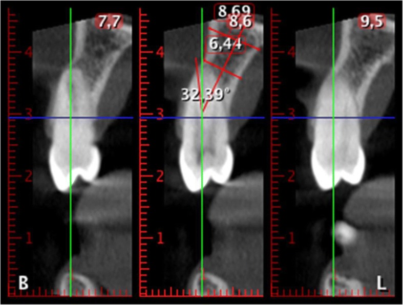
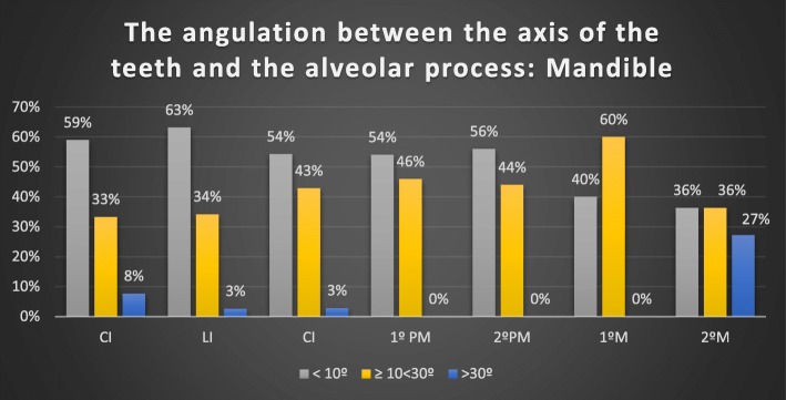
Methods: CBCT images (Planmeca ProMax 3D) of 403 teeth (208 upper teeth and 195 lower teeth) were obtained from 49 patients referred to the Dental School of Seville from January to December 2014. The thickness of the facial wall was measured at the crest, point A, 4 mm below, point B, and at the apex, point C. The second parameter was the angle formed between the dental axis and the axis of the basal bone.
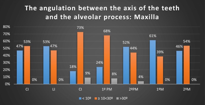
Results: A total of 403 teeth were measured. In the maxilla, 89.4% of incisors, 93.94% of canines, 78% of premolars and 70.5% of molars had a buccal bone wall thickness less than the ideal 2 mm. In the mandible, 73.5% of incisors, 49% of canines, 64% of premolars and 53% of molars had < 1 mm buccal bone thickness as measured at point B. The mean angulation in the maxilla was 11.67 ± 6.37° for incisors, 16.88 ± 7.93° for canines, 13.93 ± 8.6° for premolars, and 9.89 ± 4.8° for molars. In the mandible, the mean values were 10.63 ± 8.76° for incisors, 10.98 ± 7.36° for canines, 10.54 ± 5.82° for premolars and 16.19 ± 11.22° for molars.
Conclusions: The high incidence of a buccal wall thickness of less than 2 mm in over 80% of the assessed sites indicates the need for additional regeneration procedures, and several locations may also require custom abutments to solve the angulation problems for screw-retained crowns.
Keywords: Basal bone; Basal bone angulation; Buccal bone wall thickness; CBCT; Cone beam computed tomography; Root angulation.
Conflict of interest statement
Ethics approval and consent to participate
The ethical committee for the University of Seville approved this non-interventional study for the acquisition of the images, number 0159-N-14 (PEIBA) of the Junta de Andalucía, Spain.
Consent for publication
The authors have uploaded the Excel data file to the ‘idUS’ repository of the University of Seville.
Competing interests
The authors declare that they have no competing interests
Publisher’s Note
Springer Nature remains neutral with regard to jurisdictional claims in published maps and institutional affiliations.
Figures
References
-
- Sanz M, Cecchinato D, Ferrus J, Pjetursson EB, Lang NP, Lindhe J. A prospective, randomized-controlled clinical trial to evaluate bone preservation using implants with different geometry placed into extraction sockets in the maxilla. Clin Oral Implants Res. 2010;21(1):13–21. doi: 10.1111/j.1600-0501.2009.01824.x. - DOI - PubMed
-
- De Rouck T, Collys K, Cosyn J. Single-tooth replacement in the anterior maxilla by means of immediate implantation and provisionalization: a review. Int J Oral Maxillofac Implants. 2008;23(5):897–904. - PubMed
-
- Hammerle CH, Chen ST, Wilson TG., Jr Consensus statements and recommended clinical procedures regarding the placement of implants in extraction sockets. Int J Oral Maxillofac Implants. 2004;19(Suppl):26–28. - PubMed
-
- Mandelaris GA, Vence BS, Rosenfeld AL, Forbes DP. A classification system for crestal and radicular dentoalveolar bone phenotypes. Int J Oral Maxillofac Implants. 2013;33(3):289–296. - PubMed
MeSH terms
Substances
LinkOut - more resources
Full Text Sources
Other Literature Sources
Miscellaneous
