. 2018 Nov 21;10(468):eaat6420.
doi: 10.1126/scitranslmed.aat6420.

Two recombinant human monoclonal antibodies that protect against lethal Andes hantavirus infection in vivo

Affiliations

Two recombinant human monoclonal antibodies that protect against lethal Andes hantavirus infection in vivo

Jose L Garrido et al. Sci Transl Med. .

Abstract

Andes hantavirus (ANDV) is an etiologic agent of hantavirus cardiopulmonary syndrome (HCPS), a severe disease characterized by fever, headache, and gastrointestinal symptoms that may progress to hypotension, pulmonary failure, and cardiac shock that results in a 25 to 40% case-fatality rate. Currently, there is no specific treatment or vaccine; however, several studies have shown that the generation of neutralizing antibody (Ab) responses strongly correlates with survival from HCPS in humans. In this study, we screened 27 ANDV convalescent HCPS patient sera for their capacity to bind and neutralize ANDV in vitro. One patient who showed high neutralizing titer was selected to isolate ANDV-glycoprotein (GP) Abs. ANDV-GP-specific memory B cells were single cell sorted, and recombinant immunoglobulin G antibodies were cloned and produced. Two monoclonal Abs (mAbs), JL16 and MIB22, potently recognized ANDV-GPs and neutralized ANDV. We examined the post-exposure efficacy of these two mAbs as a monotherapy or in combination therapy in a Syrian hamster model of ANDV-induced HCPS, and both mAbs protected 100% of animals from a lethal challenge dose. These data suggest that monotherapy with mAb JL16 or MIB22, or a cocktail of both, could be an effective post-exposure treatment for patients infected with ANDV-induced HCPS.


Conflict of interest statement

Competing interests: J.L.G., F.B., and M.I.B. have a pending patent application related to this work (U.S. Provisional 62/474,681) that covers hantavirus-specific human Abs. The other authors declare that they have no competing interests.

Figures

Fig. 1.
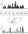
Fig. 1.. Screening of sera reactive to ANDV-GP for mAb donor identification.
Binding and neutralization assays were performed with 27 ANDV convalescent sera. (A) Binding index analysis of ANDV-GP IgG sera using a cell-based assay. 293T cells expressing ANDV-GP were incubated with a 1:1000 dilution of sera from ANDV convalescent or healthy subjects followed by detection of total IgG by flow cytometry analysis (n = 1). (B) Representative ANDV pv neutralization assay is shown, and ANDV-GP pvs were incubated with sera from ANDV convalescent subject at different dilutions (1:50 to 1:20,000) and used to infect HEK293-IB3 cells. Upon productive infection, pseudoparticles produce GFP (n = 4). (C) Representative neutralization curves of ANDV convalescent sera with high, medium, and low neutralization activities and a healthy control (C2). The y axis displays the percent pseudovirus neutralization, and the x axis displays the reciprocal of the dilution (log) (n = 4; representative experiment is shown). (D) IC50 for 27 ANDV convalescent subjects and 5 healthy controls (n = 4). The y axis displays the reciprocal of the concentration. Error bars represent the SD of the mean. P1 to P27, ANDV convalescent subjects; C1 to C5, healthy control subjects.
Fig. 2.
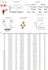
Fig. 2.. Experimental flow chart for identification and isolation of human mAbs against ANDV-GP.
(A) PBMCs isolated from one HCPS convalescent subject (P10) were used for sorting of ANDV-specific memory cells, and IGVH and IGVL were cloned in an IgG1 backbone vector. Selected mAb clones were recombinantly produced and purified, followed by characterization of binding and in vitro neutralization. FITC, fluorescein isothiocyanate. (B) List of IgG variable gene sequences analyzed using I-Blast and IMGT database shows the information for each Ab heavy and light variable chain.
Fig. 3.
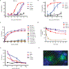
Fig. 3.. In vitro characterization of human mAbs.
(A) Neutralization assay. HEK293-IB3 cells infected with a standardized amount of ANDV-GP pv in the presence of a 10-fold dilution titration curve of purified polyclonal IgG (P10), mAb MIB22, mAb JL16, or isotype control. The percent neutralization as a measure of IgG concentrations is shown (n = 2). (B) FRNT using live ANDV (Chile-9717869). Vero E6 cells were incubated with ANDV in the presence of mAbs MIB22, mAb JL16, or isotype control (0.1 to 62.5 μg/ml), and after 7 days, viral antigen was visualized by immunoperoxidase assay. The percent neutralization as a measure of Ab concentrations is shown (n = 3). (C) Binding assay. ANDV-GP-293T cells, mock-transfected 293T cells, and VSV-G–transfected 293T cells were incubated with purified IgG (1 to 15 μg/ml) from P10, a healthy donor (C4), or mAb JL16 or MIB22. Samples were analyzed by flow cytometry (n = 2 to 3; representative experiment is shown). (D) mAb off-rate determination. ANDV-GP-293T cells were incubated with purified polyclonal IgG (15 μg/ml) from a control donor (C4), ANDV convalescent patient (P10), or mAb JL16 or MIB22 at 4°C; after the indicated times at 37°C, surface levels were detected using an Alexa Fluor 488 anti-human IgG Ab and flow cytometry. The percent of normalized staining with respect to time = 0 is shown (n = 3). (E) ELISA using ANDV-GP pv as the coating antigen and purified polyclonal IgG (P10) (0.001 to 10 μg/ml), isotype control Ab, or mAbs JL16 and MIB22 (n = 3). OD, optical density. (F) Confocal imaging. Surface staining of ANDV-GP-293T cells that were incubated with MIB22 mAb at 1 μg/ml followed by staining with an anti-human IgG conjugated with Alexa Fluor 488. 4′,6-Diamidino-2-phenylindole (DAPI) nuclear staining dye was used, and cells were visualized using confocal microscopy (n = 4; representative experiment is shown). Error bars represent the SD of the mean.
Fig. 4.
Fig. 4.. Passive transfer of mAb protects against ANDV infection.
Twenty-four hamsters were inoculated intranasally with 200 FFUs of ANDV. (A) Groups of six hamsters were then administered MIB22, JL16, cocktail, or isotype control (50 mg/kg) intraperitoneally at days 3 and 8 after inoculation. Hamsters were monitored for disease. Survival was statistically evaluated using log-rank (Mantel-Cox) tests (P < 0.0001). (B) ANDV-N ELISA from sera collected from surviving hamsters on 36 DPI for evidence of ANDV infection (n = 18). (C) Animals that survived to 36 DPI were euthanized, and ANDV-specific S-segment RNA was quantified using qRT-PCR in the lung tissue (n = 18).

References

    1. Hjelle B, Torres-Pérez F, Hantaviruses in the Americas and their role as emerging pathogens. Viruses 2, 2559–2586 (2010). - PMC - PubMed
    1. Vaheri A, Strandin T, Hepojoki J, Sironen T, Henttonen H, Makela S, Mustonen J, Uncovering the mysteries of hantavirus infections. Nat. Rev. Microbiol. 11, 539–550 (2013). - PubMed
    1. Martinez VP, Bellomo C, San Juan J, Pinna D, Forlenza R, Elder M, Padula PJ, Person-to-person transmission of Andes virus. Emerg. Infect. Dis. 11, 1848–1853 (2005). - PMC - PubMed
    1. Macneil A, Nichol ST, Spiropoulou CF, Hantavirus pulmonary syndrome. Virus Res. 162, 138–147 (2011). - PubMed
    1. Figueiredo LT, Souza WM, Ferres M, Enria DA, Hantaviruses and cardiopulmonary syndrome in South America. Virus Res. 187, 43–54 (2014). - PubMed

Publication types

MeSH terms