. 2019 Jan 25;294(4):1104-1125.
doi: 10.1074/jbc.RA118.006072. Epub 2018 Nov 21.

CRISPR/Cas9 and glycomics tools for Toxoplasma glycobiology

Affiliations

CRISPR/Cas9 and glycomics tools for Toxoplasma glycobiology

Elisabet Gas-Pascual et al. J Biol Chem. .

Abstract

Infection with the protozoan parasite Toxoplasma gondii is a major health risk owing to birth defects, its chronic nature, ability to reactivate to cause blindness and encephalitis, and high prevalence in human populations. Unlike most eukaryotes, Toxoplasma propagates in intracellular parasitophorous vacuoles, but like nearly all other eukaryotes, Toxoplasma glycosylates many cellular proteins and lipids and assembles polysaccharides. Toxoplasma glycans resemble those of other eukaryotes, but species-specific variations have prohibited deeper investigations into their roles in parasite biology and virulence. The Toxoplasma genome encodes a suite of likely glycogenes expected to assemble N-glycans, O-glycans, a C-glycan, GPI-anchors, and polysaccharides, along with their precursors and membrane transporters. To investigate the roles of specific glycans in Toxoplasma, here we coupled genetic and glycomics approaches to map the connections between 67 glycogenes, their enzyme products, the glycans to which they contribute, and cellular functions. We applied a double-CRISPR/Cas9 strategy, in which two guide RNAs promote replacement of a candidate gene with a resistance gene; adapted MS-based glycomics workflows to test for effects on glycan formation; and infected fibroblast monolayers to assess cellular effects. By editing 17 glycogenes, we discovered novel Glc0-2-Man6-GlcNAc2-type N-glycans, a novel HexNAc-GalNAc-mucin-type O-glycan, and Tn-antigen; identified the glycosyltransferases for assembling novel nuclear O-Fuc-type and cell surface Glc-Fuc-type O-glycans; and showed that they are important for in vitro growth. The guide sequences, editing constructs, and mutant strains are freely available to researchers to investigate the roles of glycans in their favorite biological processes.

Keywords: CRISPR/Cas; Toxoplasma gondii; glycan; glycobiology; glycogene; glycomics; glycosyltransferase; mass spectrometry (MS); parasitology; protein glycosylation.


Conflict of interest statement

The authors declare that they have no conflicts of interest with the contents of this article

Figures

Figure 1.
Figure 1.
Fitness of glycogenes. Fitness data for glycogenes for invasion of and growth in human fibroblasts were extracted from Ref. and graphed according to rank order of fitness value for separate categories of glycosylation as indicated at the bottom. Fitness scores are based on a log2 scale, and values below −2.5 (red dashed line) have increasing likelihood of being essential. Filled data points represent genes for which mutants are available: from this study (green) or from other studies (blue). Red filled circles, unsuccessful attempts from this study; unfilled circles, no mutants attempted. Genes for which pDGs are available or that have been mutated elsewhere are labeled in black; others are labeled in gray. Gene names correspond to listings in Table 1.
Figure 2.
Figure 2.
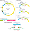
Plasmids and gene replacement. The previously described pU6-Universal plasmid (A), designed for single-site CRISPR/Cas9-mediated editing (26), contains a BsaI site for convenient introduction of a synthetic duplex guide DNA (B). pU6 was modified by the addition of an XhoI site at the 5′-end of its guide DNA cassette to generate p2 (C) or at its 3′-end to generate p3 (D). Thus, the guide 2 cassette of p2 could be conveniently excised from and religated into p3 to generate the dual-guide plasmid pDG (E). F, gene replacement strategy. Co-transient transfection of pDG, expressing the gRNAs and Cas9, with the DHFR amplicon, results in the excision of the intervening genomic DNA (a) and ligation of the DHFR amplicon by nonhomologous end joining (b). The desired replacement clones are selected in the presence of pyrimethamine (c) and screened by PCR using gene-specific primers (GSP) flanking the excision sites.
Figure 3.
Figure 3.
Phenotype analysis. Clones were analyzed using a plaque assay on confluent HFF monolayers, and plaque areas were measured 6 days after infection. A, image comparing plaques from RH (parental) and alg8Δ strains. B, dot plots from pairwise comparisons of mutant and RH strains. Gene names are color-coded to indicate their glycosylation pathway association (red, N-glycans; green, other secretory pathway glycans; purple, cytoplasmic glycans; black, precursor assembly), and fitness scores from Fig. 1 are indicated in parentheses. To facilitate comparisons between trials, the mean areas of RH plaques were normalized to 1, and the respective mutant plaque areas were scaled accordingly (only one representative RH experiment is graphed for clarity). Mean values ± 2 S.D. values (95% confidence interval) are shown with red bars. Statistical significance is shown at the top. Data were pooled from three independent trials, which each independently conformed to the same trend.
Figure 4.
Figure 4.
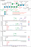
N-Glycan glycogenes and nano-LC-MS analysis of parasite N-glycans. A, the N-glycan precursor is assembled on dolichol-pyrophosphate, one sugar at a time from right to left. Genes that contribute to biosynthetic steps are depicted. The status of gene editing is color-coded according to the key. The complete Glc3Man6GlcNAc2 glycan is transferred en bloc to protein Asn side chains in an NX(S/T) motif by stt3 and variably trimmed by α-glucosidase-I and -II (Table 1A). Glycans are represented according to the Consortium for Functional Glycomics system of nomenclature (57). B and C, N-glycans were prepared by treating tryptic digests of total parasite protein powders with PNGase F. The released glycans were permethylated and subjected to nLC on a C18 column followed by MSn in a linear ion-trap MS. Extracted ion chromatograms for all detected N-glycan–like, lithiated (Li+) composition (H1N2–H8N2) species are shown before and after digestion with jack bean α-mannosidase (α1–2/3/6) or A. satoi (α1–2) mannosidase to probe the identity of the hexose (H) species. The two peaks observed for each species are α- and β-anomers. Minor related N-glycan-like species are shown in Fig. S6A. B, RH (parental). C, alg5Δ mutant, which is unable to form Dol-P-Glc, the Glc donor for terminal α-glucosylation.
Figure 5.
Figure 5.
O-Glycan, C-mannose, and polysaccharide glycogenes in the secretory pathway and their glycomic analysis. A, mucin-type O-glycans. Shown are Tn antigen (GalNAcα-Ser/Thr) and the N2 disaccharide, consisting of a HexNAc of uncertain identity (potentially GalNAc) linked to αGalNAc, inferred from MS glycomic studies and metabolic and lectin labeling studies (see “Results and discussion”). Five pp-αGalNAcTs are predicted to catalyze formation of the Tn antigen, and T2 and T3 are confirmed. T2 is required for accumulation of N2, but the enzyme responsible for the second sugar is unknown, except that genes in green in D are not required. B, TSR-type Glc-Fuc- disaccharide and its documented GT genes (see Fig. 7C). C, GT for C-mannosylation. D, four genes encoding predicted sugar nucleotide-dependent GT that appear to be type 2 membrane proteins in the secretory pathway. The glycans to which they contribute are yet to be identified. E, GT for assembly of the oocyst wall β3-glucan, whose priming mechanism is unknown. F–H, O-glycans were released by reductive β-elimination, permethylated, and subjected to nLC on a C18 column followed by MSn in a linear ion-trap MS as in Fig. 4. Extracted ion chromatograms for N2 (drawn as GalNAc-GalNAc-) and HdH (drawn as Glc-Fuc-) species (lithiated adducts ±70 ppm) and the DP4 standard are shown. F, parental strain (type 2 Pru) and two derivative mutant strains (pp-αGalNAcT2 and pp-αGalNAcT3). G, the parental strain (type 1 RH) and the derivative galEΔ strain and an extract of host hTERT cells showing HN, HNSa (Sa = sialic acid), and HdH, but lacking N2. H, RH and two derivative mutants predicted to encode the sequential addition of αFuc (POFUT2Δ) and βGlc (glt2Δ or β3GlcTΔ). I, Western blot analysis of total RH strain tachyzoites for the Tn-antigen, probed with mAb 5F4. The Coomassie Blue–stained gel confirms equal sample loading. Similar results were obtained with mAb 1E3, except that mAb-reactive proteins were differentially emphasized.
Figure 6.
Figure 6.
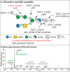
GIPL and GPI-anchor biosynthesis glycogenes. A, free GIPLs and GPI-anchors share a related glycan linked to a diacylglycerol on the right. Glycan assembly proceeds stepwise from right to left along the upper arm by the enzymes predicted, followed by the lower arm (58). The GPI-anchor precursor may receive the lower arm β4GalNAc, whereas the GIPL may also receive an α4Glc (dashed line). Phosphoethanolamine is then conjugated to the terminal α2Man, and the resulting NH2 group of the GPI can be conjugated to the polypeptide C terminus by a multisubunit transamidase. Potential fatty acyl modifications are not shown. B, the glycan region of GPI-anchored proteins enriched from RH strain tachyzoites was released by HF (after saponification), which cleaves phosphodiester bonds, subjected to re-N-acetylation and permethylation, and analyzed by MSn. m/z values corresponding to glycans containing one or two of the sugars of the predicted lower arm are indicated in gray, but were not detected in this study.
Figure 7.
Figure 7.
Cytoplasmic GT glycogenes. A, assembly of the hydroxyproline-linked pentasaccharide on the E3SCFUb-ligase subunit Skp1 occurs stepwise from right to left. B, formation of O-Fuc on Ser/Thr residues of nucleocytoplasmic proteins. C, AAL (green) binding to four nuclei of parental RH strain parasites (white arrow, top left), housed within the same parasitophorous vacuole within a host fibroblast, is lost in the six spyΔ cells shown (bottom left). To the right, IMC3 (red) labeling localizes to the parasite inner membrane complex whose organization varies according to the cell cycle. D, AAL labeling of Western blots of parental strain whole-cell extracts is lost in spyΔ cells. Subjecting the blotted membrane to conditions of β-elimination removed the majority of labeling, consistent with its O-linkage. E, genes associated with amylopectin assembly. Genes associated with its disassembly are shown in Table 1I. F, other genes predicted to encode sugar nucleotide–dependent cytoplasmic (or nuclear) GTs.
Figure 8.
Figure 8.
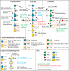
Precursor assembly pathways and transporters. Predicted and validated glycogenes associated with the formation and transport of sugar nucleotides, and formation of dolichol-linked monosaccharides, that are utilized as sugar donors by glycosyltransferases. A, UDP-GlcNAc and UDP-GalNAc are derived from Glc via Glc6P. B, UDP-Glc, UDP-Gal, and Dol-P-Glc can derive from either Glc or Gal. C, GDP-Man, Dol-P-Man, and GDP-Fuc also ultimately derive from Glc. D, destination glycans for the precursors boxed in orange in A–C. E, transporters transfer sugar nucleotides from the cytoplasm where they are synthesized into the lumina of vesicles containing the glycosyltransferases. The scramblases that reorient Dol-P-Glc and Dol-P-Man from the cytoplasmic to the luminal face of the rER are unknown. F, definition of symbols and color coding of the status of gene analyses.

References

    1. Kim K., and Weiss L. M. (2004) Toxoplasma gondii: the model apicomplexan. Int. J. Parasitol. 34, 423–432 10.1016/j.ijpara.2003.12.009 - DOI - PMC - PubMed
    1. Pappas G., Roussos N., and Falagas M. E. (2009) Toxoplasmosis snapshots: global status of Toxoplasma gondii seroprevalence and implications for pregnancy and congenital toxoplasmosis. Int. J. Parasitol. 39, 1385–1394 10.1016/j.ijpara.2009.04.003 - DOI - PubMed
    1. Pereira-Chioccola V. L., Vidal J. E., and Su C. (2009) Toxoplasma gondii infection and cerebral toxoplasmosis in HIV-infected patients. Future Microbiol. 4, 1363–1379 10.2217/fmb.09.89 - DOI - PubMed
    1. Luft B. J., and Remington J. S. (1992) Toxoplasmic encephalitis in AIDS. Clin. Infect. Dis. 15, 211–222 10.1093/clinids/15.2.211 - DOI - PubMed
    1. Lambert H., and Barragan A. (2010) Modelling parasite dissemination: host cell subversion and immune evasion by Toxoplasma gondii. Cell. Microbiol. 12, 292–300 10.1111/j.1462-5822.2009.01417.x - DOI - PubMed

Publication types

Substances