. 2018 Dec 28;293(52):19974-19981.
doi: 10.1074/jbc.AC118.005771. Epub 2018 Nov 21.

Establishment of a continuous in vitro culture of Babesia duncani in human erythrocytes reveals unusually high tolerance to recommended therapies

Affiliations

Establishment of a continuous in vitro culture of Babesia duncani in human erythrocytes reveals unusually high tolerance to recommended therapies

Amanah Abraham et al. J Biol Chem. .

Abstract

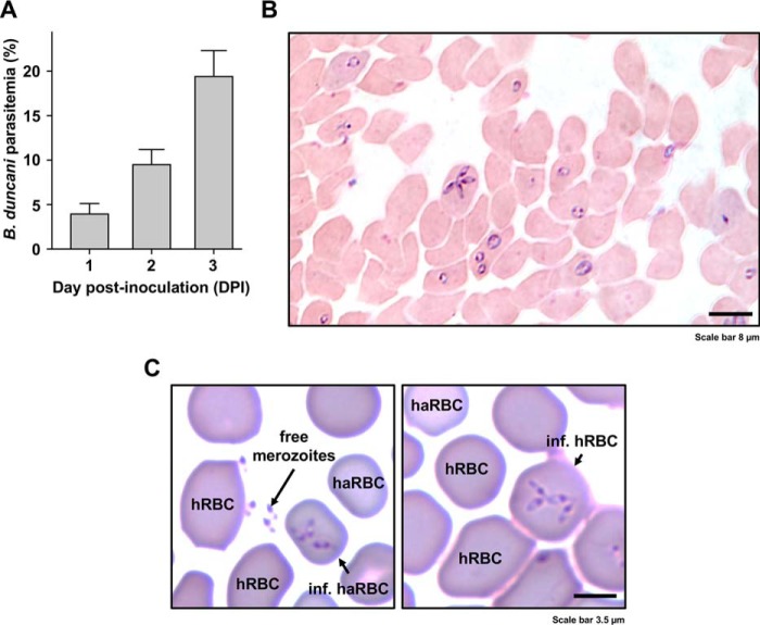
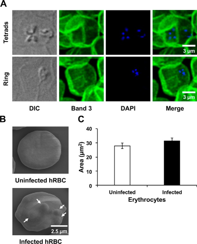
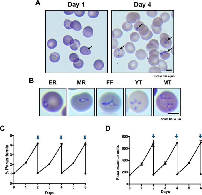
Human babesiosis is an emerging tick-borne disease caused by apicomplexan parasites of the genus Babesia Clinical cases caused by Babesia duncani have been associated with high parasite burden, severe pathology, and death. In both mice and hamsters, the parasite causes uncontrolled fulminant infections, which ultimately lead to death. Resolving these infections requires knowledge of B. duncani biology, virulence, and susceptibility to anti-infectives, but little is known and further research is hindered by a lack of relevant model systems. Here, we report the first continuous in vitro culture of B. duncani in human red blood cells. We show that during its asexual cycle within human erythrocytes, B. duncani develops and divides to form four daughter parasites with parasitemia doubling every ∼22 h. Using this in vitro culture assay, we found that B. duncani has low susceptibility to the four drugs recommended for treatment of human babesiosis, atovaquone, azithromycin, clindamycin, and quinine, with IC50 values ranging between 500 nm and 20 μm These data suggest that current practices are of limited effect in treating the disease. We anticipate this new disease model will set the stage for a better understanding of the biology of this parasite and will help guide better therapeutic strategies to treat B. duncani-associated babesiosis.

Keywords: Babesia duncani; apicomplexa; cell culture; erythrocyte; human babesiosis; in vitro culture; infectious disease; inhibitor; parasitology; red blood cells; tick-borne disease.


Conflict of interest statement

The authors declare that they have no conflicts of interest with the contents of this article. The content is solely the responsibility of the authors and does not necessarily represent the official views of the National Institutes of Health.

Figures

Figure 1.
Figure 1.
Continuous in vitro culture of B. duncani in hamster RBCs and transfer to human RBCs. A, parasitemia expressed as the percentage of hamster RBCs infected by the parasite at 1, 2, and 3 days post-inoculation (DPI) in a representative experiment. Columns represent mean ± S.E. (error bars) of six biological replicates. B, micrograph of the intracellular development of cultured B. duncani in hamster RBCs in a Giemsa-stained smear prepared at 3 days post-inoculation. The parasitemia was 15% in this sample. C, transfer of B. duncani to human RBCs. B. duncani-infected hamster RBCs (haRBCs) were freshly harvested and maintained in culture in the presence of human RBCs (hRBCs). Left, free merozoites and infected haRBCs. Right, successful development of B. duncani in hRBCs. Human RBCs are distinguishable from haRBCs by their larger size and darker staining.
Figure 2.
Figure 2.
Confocal and scanning EM imaging of B. duncani-infected human RBCs. A, infected human RBCs were fixed and incubated with anti-Band3 mouse mAb (secondary antibody coupled to Alexa Fluor 488). Parasite DNA was stained with DAPI. B, representative images of uninfected and B. duncani-infected human RBCs visualized by scanning EM. Protrusions caused by the parasite are indicated by white arrows. C, quantification of the size of uninfected and B. duncani-infected human red blood cells from images collected by scanning EM. Data represent mean values of 20 uninfected and 19 infected erythrocytes. Error bars, S.E. The area of the examined erythrocytes was determined using Fiji. DIC, differential interference contrast.
Figure 3.
Figure 3.
Continuous in vitro culture of B. duncani in human RBCs. A, representative images of Giemsa-stained blood smears of infected human RBCs at 1% parasitemia on day 1 and 8% parasitemia on day 4. B, representative images of the various stages identified in blood smears of B. duncani-infected human RBCs. EE, early rings; MR, mature rings; FF, filamentous forms; YT, young tetrads; MT, mature tetrads. C and D, growth of B. duncani over a 6-day period in human RBCs with culture dilution at day 2. Arrows indicate when cultures were diluted to 1% parasitemia. Parasitemia was determined by light microscopy and counting of 3,000–5,000 RBCs (C) or the SYBR Green I assay (D). Data represent mean values measured in triplicates. Error bars, S.D.
Figure 4.
Figure 4.
Evaluation of the in vitro drug susceptibility of B. duncani to atovaquone (ATV) (A), azithromycin (AZ) (B), clindamycin (CLI) (C), and quinine (QUI) (D) at concentrations up to 100 μm. Parasitemia was determined by the SYBR Green I assay at 60 h after the addition of the drugs. Wells with untreated infected erythrocytes and 0.1% DMSO vehicle were set as 100% growth, and wells with compounds at 100 μm in 0.1% DMSO were set as 0% growth. Each experiment was performed in triplicate, and data presented represent one of two biological replicates. GraphPad Prism (version 7.0a) was used to generate sigmoidal dose–response curves by fitting a nonlinear regression curve to the data. Each data point represents mean value measured in triplicates with error bars indicating S.D.

Similar articles

Cited by

References

    1. Gelfand J. A., and Callahan M. V. (2003) Babesiosis: an update on epidemiology and treatment. Curr. Infect. Dis. Rep. 5, 53–58 10.1007/s11908-003-0065-z - DOI - PubMed
    1. Conrad P. A., Kjemtrup A. M., Carreno R. A., Thomford J., Wainwright K., Eberhard M., Quick R., Telford S. R. 3rd, and Herwaldt B. L. (2006) Description of Babesia duncani n. sp. (Apicomplexa: Babesiidae) from humans and its differentiation from other piroplasms. Int. J. Parasitol. 36, 779–789 10.1016/j.ijpara.2006.03.008 - DOI - PubMed
    1. Scott J. D., and Scott C. M. (2018) Human babesiosis caused by Babesia duncani has widespread distribution across Canada. Healthcare (Basel) 6, E49 10.3390/healthcare6020049 - DOI - PMC - PubMed
    1. Leiby D. A. (2011) Transfusion-transmitted Babesia spp.: bull's-eye on Babesia microti. Clin. Microbiol. Rev. 24, 14–28 10.1128/CMR.00022-10 - DOI - PMC - PubMed
    1. Zamoto-Niikura A., Tsuji M., Imaoka K., Kimura M., Morikawa S., Holman P. J., Hirata H., and Ishihara C. (2014) Sika deer carrying Babesia parasites closely related to B. divergens, Japan. Emerg. Infect. Dis. 20, 1398–1400 10.3201/eid2008.130061 - DOI - PMC - PubMed

Publication types

MeSH terms

LinkOut - more resources