. 2018 Dec 11;115(50):12781-12786.
doi: 10.1073/pnas.1803826115. Epub 2018 Nov 21.

The unusual rainbow trout sex determination gene hijacked the canonical vertebrate gonadal differentiation pathway

Affiliations

The unusual rainbow trout sex determination gene hijacked the canonical vertebrate gonadal differentiation pathway

Sylvain Bertho et al. Proc Natl Acad Sci U S A. .

Abstract

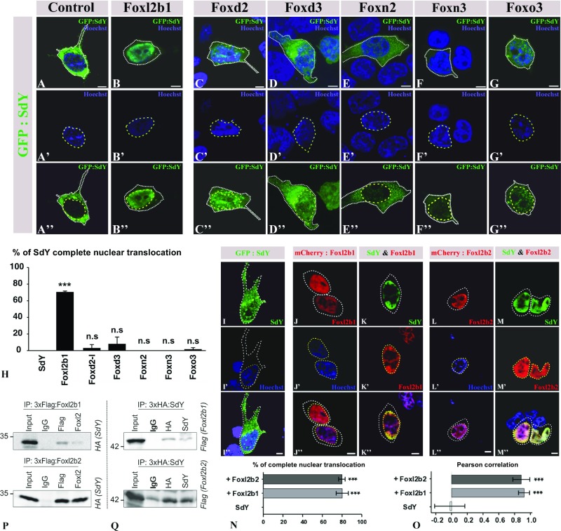
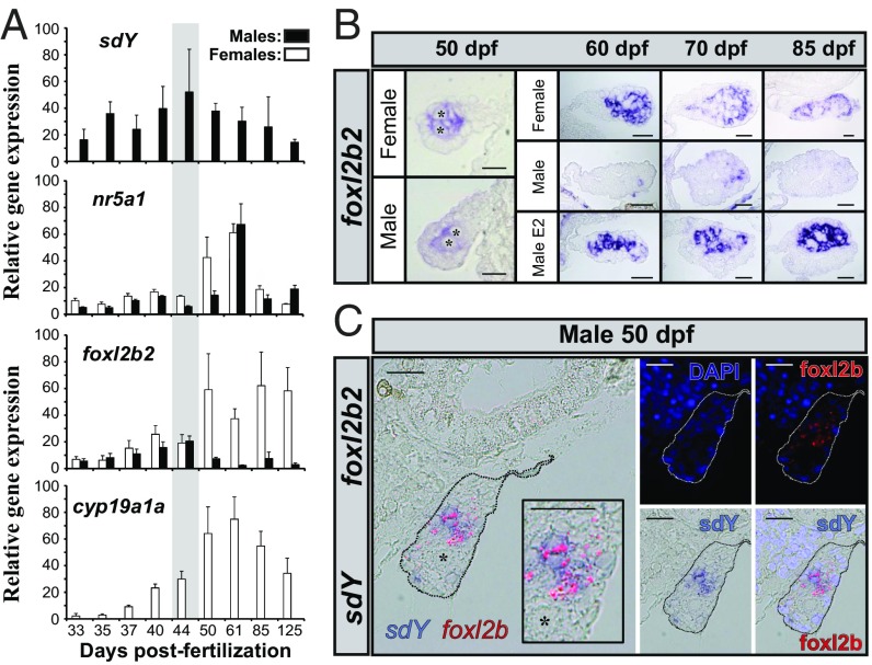
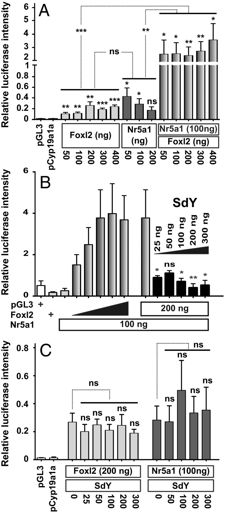
Evolutionary novelties require rewiring of transcriptional networks and/or the evolution of new gene functions. Sex determination (SD), one of the most plastic evolutionary processes, requires such novelties. Studies on the evolution of vertebrate SD revealed that new master SD genes are generally recruited from genes involved in the downstream SD regulatory genetic network. Only a single exception to this rule is currently known in vertebrates: the intriguing case of the salmonid master SD gene (sdY), which arose from duplication of an immune-related gene. This exception immediately posed the question of how a gene outside from the classical sex differentiation cascade could acquire its function as a male SD gene. Here we show that SdY became integrated in the classical vertebrate sex differentiation cascade by interacting with the Forkhead box domain of the female-determining transcription factor, Foxl2. In the presence of Foxl2, SdY is translocated to the nucleus where the SdY:Foxl2 complex prevents activation of the aromatase (cyp19a1a) promoter in cooperation with Nr5a1 (Sf1). Hence, by blocking a positive loop of regulation needed for the synthesis of estrogens in the early differentiating gonad, SdY disrupts a preset female differentiation pathway, consequently allowing testicular differentiation to proceed. These results also suggest that the evolution of unusual vertebrate master sex determination genes recruited from outside the classical pathway like sdY is strongly constrained by their ability to interact with the canonical gonadal differentiation pathway.

Keywords: Forkhead box proteins; evolution; fish; sex determination; sex differentiation.


Conflict of interest statement

The authors declare no conflict of interest.

Figures

Fig. 1.
Fig. 1.
SdY conserves the structure of the IRF protein–protein interaction domain and interacts with the Forkhead box domain of Fox proteins. (A) SdY shares structural homologies with IAD, a protein–protein interaction domain. The structure of SdY (in gray) was modeled using the crystal structure of IRF5 as a template (in green). This SdY structure reveals eight β-sheets forming a β-sandwich and three α-helices that are highly conserved with IRF5. (B) SdY interacts in yeast with Fox proteins through their highly conserved DNA-binding domain. The alignments of the SdY-Fox interacting clone sequences (gray lines) delineate the minimum domain or selected interacting domain needed for an effective interaction with SdY in yeast, which is the Forkhead box domain (110 aa, black lines). The 11 Fox proteins characterized in the Y2H screen are represented by open cylinders with numbers of interacting clones indicated on the right side.
Fig. 2.
Fig. 2.
SdY interacts with Foxl2, resulting in its nuclear translocation. (AH) GFP:SdY alone (AA″) and GFP:SdY in combination with different trout Fox proteins, Foxl2 (Foxl2b1) (BB″), Foxd2 (CC″), Foxd3 (DD″), Foxn2 (EE″), Foxn3 (FF″), Foxo3 (GG″), were cotransfected in HEK 293T cells (delimited by white dotted lines). GFP:SdY is translocated in the nucleus (delimited by yellow dotted lines and stained in blue with Hoechst staining) only in the presence of Foxl2 (BB″). (Scale bar, 5 μm.) (H) Percentage of transfected cells (mean ± standard deviation on 200 cells) in which SdY is completely translocated in the nucleus after three independent cotransfection experiments with different trout Fox proteins. Significant differences compared with SdY alone were calculated using an unpaired two-tailed Student’s t test, ***P < 0.001; ns, nonsignificant. (IO) Foxl2b1 and Foxl2b2 are both able to drive SdY complete nuclear translocation (delimited by yellow dotted lines and stained in blue with Hoechst staining). Confocal images of HEK 293T cells (delimited by white dotted lines) transiently transfected with sdY (II″), mCherry:Foxl2b1 alone (JJ″), SdY and mCherry:Foxl2b1 (KK″), mCherry:Foxl2b2 alone (LL″), SdY and mCherry:Foxl2b2 (MM″). (Scale bar, 10 µm.) (N) Quantitative analysis in the presence or absence of Foxl2b1 and Foxl2b2. Percentage of complete SdY nuclear translocation (mean ± standard deviation on 100 cells) after three independent cotransfection experiments. Statistical significances compared with SdY alone were calculated using an unpaired two-tailed Student’s t test. (O) SdY colocalizes with Foxl2 in the nucleus. SdY, SdY-Foxl2b1, and SdY-Foxl2b2 colocalizations were measured in the nucleus for SdY (n = 5), SdY and Foxl2b1 (n = 5), and SdY and Foxl2b2 (n = 5) with Pearson’s correlation. Statistical significance was calculated using an unpaired two-tailed Student’s t test, ***P < 0.001. (P and Q) SdY binds with Foxl2 in co-immunoprecipitation (IP) experiments. HEK 293T cells were transiently transfected with expression plasmids for SdY fused to a hemagglutinin tag (3xHA:SdY) and for Foxl2 fused to a 3xFlag tag (3xFlag:Foxl2b1 or 3xFlag:Foxl2b2). Whole-cell lysates were used for immunoprecipitation with anti-Flag or anti-Foxl2 (P) and with anti-HA or anti-SdY (Q) followed by immunoblotting with the appropriate antibodies. Input represents 10% whole-cell lysate. IgG mouse antibody was used as the control. In P, 3xFlag:Foxl2b1 or 3xFlag:Foxl2b2 was immunoprecipitated with either Flag (Top) or FoxL2 (Bottom) antibodies followed by immunoblotting with an antibody against the HA tag to reveal the interaction with 3xHA:SdY (SdY). In Q, 3xHA:SdY was immunoprecipitated with an HA or SdY antibody, followed by immunoblotting with an antibody against the Flag tag to reveal 3xFlag:Foxl2b1 (Foxl2b1) (Top) or 3xFlag:Foxl2b2 (Foxl2b2) (Bottom).
Fig. 3.
Fig. 3.
The gonadal expression patterns of sdY, foxl2, nr5a1, and cyp19a1a (A and B) are in agreement with a repressive effect of SdY on the Foxl2 positive regulation of the aromatase promoter (C). (A) Gene expression profiles of sdY, nr5a1, foxl2b2, and cyp19a1a in male and female gonads from 33 to 125 dpf. All values represent the mean ± standard deviation of three biological replicates (in percentage of the highest measured value). The gray area highlights the period before the sexually dimorphic expression of trout foxl2 genes during gonadal differentiation. (B) Gonadal localization of foxl2b2 transcripts (NBT/BCIP signal in blue) in male and female gonads. foxl2b2 is expressed in somatic cells of both female and male gonads at 50 dpf and only in female gonads at later stages (60, 70, and 85 dpf). In males fed with estrogens (male E2) foxl2b2 is strongly up-regulated compared with control males quickly after (60 dpf) the application of the treatment (55 dpf). (Scale bar, 20 µm.) (C) Colocalization by double in situ hybridization of sdY (NBT/BCIP signal in blue) and foxl2b2 (HNPP/Fast Red signal in red fluorescence) in somatic cells of a rainbow trout male differentiating gonad at 50 dpf. Cell nuclei are shown in the dark-field panels stained with DAPI either with or without the HNPP fluorescent detection of foxl2b2. Germ cells are shown by an asterisk. (Scale bar, 20 µm.)
Fig. 4.
Fig. 4.
SdY prevents the Foxl2/Nr5a1 positive regulation of the cyp19a1a promoter. Medaka cyp19a1a promoter activity (pGL3-cyp19a1a promoter coupled to a firefly luciferase) was measured using a luciferase reporter assay after HEK 293T cell cotransfection with either medaka nr5a1 and/or foxl2 expression plasmids and variable concentrations of the rainbow trout sdY expression plasmid. All results were calculated as the mean ± SEM of three biological replicates in two independent experiments. (A) Foxl2 and Nr5a1 act in synergy to induce cyp19a1a expression. Medaka cyp19a1a luciferase assay with variable concentrations of foxl2 (50–400 ng) and nr5a1 (50–200 ng) or 100 ng of nr5a1 with variable concentrations of foxl2 (50–400 ng). Statistical significances of luciferase activity within treatments were tested using an unpaired two-tailed Student’s t test. Effects of foxl2 alone and nr5a1 alone compared with their synergistic effect (shown by asterisks on the dotted lines joining the different groups) were tested by a one-way ANOVA with a post hoc Dunnett test. (B) SdY prevents Foxl2/Nr5a1 positive regulation of the cyp19a1a promoter. Medaka cyp19a1a luciferase assay with variable concentrations of foxl2 (50–400 ng) combined with a fixed concentration of nr5a1 (100 ng) and a fixed concentration of foxl2 (200 ng) and nr5a1 (100 ng) combined with variable concentrations of sdY (25–300 ng). Empty plasmid control (pGL3) and Foxl2 alone (200 ng) are depicted by a + sign. Statistical significances of luciferase activity were tested using a Mann–Whitney U test. (C) SdY does not repress cyp19a1a promoter expression induced by medaka Foxl2 alone or Nr5a1 alone. Medaka cyp19a1a luciferase assay with fixed foxl2 (200 ng) or nr5a1 (100 ng) concentrations combined with variable concentrations of sdY (25–300 ng). Statistical significances of luciferase activity within treatments were tested using an unpaired two-tailed Student’s t test. The effect of foxl2 or nr5a1 alone compared with foxl2 or nr5a1 and variable concentrations of sdY was tested by a one-way ANOVA with post hoc Tukey tests. ***P < 0.001; **P < 0.01; *P < 0.1; ns, P > 0.05 (nonsignificant).

Similar articles

Cited by

References

    1. Herpin A, Schartl M. Plasticity of gene-regulatory networks controlling sex determination: Of masters, slaves, usual suspects, newcomers, and usurpators. EMBO Rep. 2015;16:1260–1274. - PMC - PubMed
    1. Graves JAM. How to evolve new vertebrate sex determining genes. Dev Dyn. 2013;242:354–359. - PubMed
    1. Lin Y-T, Capel B. Cell fate commitment during mammalian sex determination. Curr Opin Genet Dev. 2015;32:144–152. - PMC - PubMed
    1. Windley SP, Wilhelm D. Signaling pathways involved in mammalian sex determination and gonad development. Sex Dev. 2015;9:297–315. - PubMed
    1. Kikuchi K, Hamaguchi S. Novel sex-determining genes in fish and sex chromosome evolution. Dev Dyn. 2013;242:339–353. - PubMed

Publication types