. 2019 Feb 5;93(4):e01861-18.
doi: 10.1128/JVI.01861-18. Print 2019 Feb 15.

Packaging of the Influenza Virus Genome Is Governed by a Plastic Network of RNA- and Nucleoprotein-Mediated Interactions

Affiliations

Packaging of the Influenza Virus Genome Is Governed by a Plastic Network of RNA- and Nucleoprotein-Mediated Interactions

Hardin Bolte et al. J Virol. .

Abstract

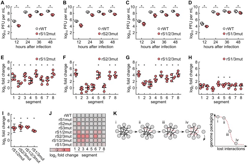
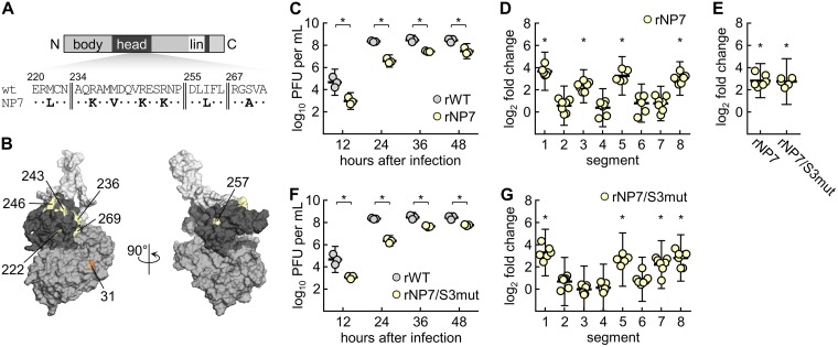
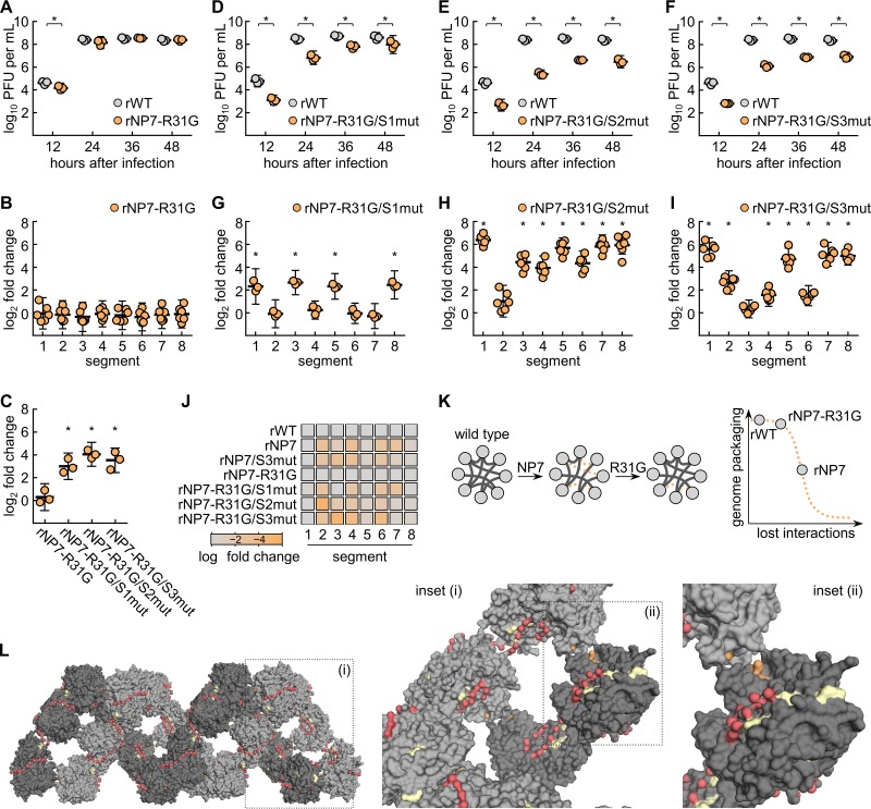
The genome of influenza A virus is organized into eight ribonucleoproteins, each composed of a distinct RNA segment bound by the viral polymerase and oligomeric viral nucleoprotein. Packaging sequences unique to each RNA segment together with specific nucleoprotein amino acids are thought to ensure the precise incorporation of these eight ribonucleoproteins into single virus particles, and yet the underlying interaction network remains largely unexplored. We show here that the genome packaging mechanism of an H7N7 subtype influenza A virus widely tolerates the mutation of individual packaging sequences in three different RNA segments. However, combinations of these modified RNA segments cause distinct genome packaging defects, marked by the absence of specific RNA segment subsets from the viral particles. Furthermore, we find that combining a single mutated packaging sequence with sets of specific nucleoprotein amino acid substitutions greatly impairs the viral genome packaging process. Along with previous reports, our data propose that influenza A virus uses a redundant and plastic network of RNA-RNA and potentially RNA-nucleoprotein interactions to coordinately incorporate its segmented genome into virions.IMPORTANCE The genome of influenza A virus is organized into eight viral ribonucleoproteins (vRNPs); this provides evolutionary advantages but complicates genome packaging. Although it has been shown that RNA packaging sequences and specific amino acids in the viral nucleoprotein (NP), both components of each vRNP, ensure selective packaging of one copy of each vRNP per virus particle, the required RNA-RNA and RNA-NP interactions remain largely elusive. We identified that the genome packaging mechanism tolerates the mutation of certain individual RNA packaging sequences, while their combined mutation provokes distinct genome packaging defects. Moreover, we found that seven specific amino acid substitutions in NP impair the function of RNA packaging sequences and that this defect is partially restored by another NP amino acid change. Collectively, our data indicate that packaging of the influenza A virus genome is controlled by a redundant and plastic network of RNA/protein interactions, which may facilitate natural reassortment processes.

Keywords: complexity; influenza; nucleoprotein; packaging; vRNPs.


Figures

FIG 1
FIG 1
SC35M viruses with a single mutated packaging sequence show no or minor genome packaging defects. (A to C) Schematic representation of the SC35M vRNA segments (S) 1, 2, and 3. Wild-type and mutated nucleotide sequences are shown in black and red, respectively, and numbered in the positive sense. (D to F) Multicycle replication kinetics of the indicated mutant viruses compared to wild-type SC35M (rWT) (n = 3 to 5). (G to I) Relative quantification of the vRNA segment levels between mutant and wild-type virus particles by RT-qPCR. RNA levels were normalized to identical PFU numbers (n = 3 to 5). (J) Relative quantification of HAU-to-PFU ratios between mutant and wild-type virus particles (n = 3 to 5). In panels D to J, data are presented as means ± 95% confidence intervals. *, P < 0.05 (Student t test).
FIG 2
FIG 2
SC35M viruses with multiple mutated packaging sequences in different vRNA segments show distinct genome packaging defects. (A to D) Multicycle replication kinetics of the indicated mutant viruses compared to wild-type SC35M (rWT) (n = 3). (E to H) Relative quantification of the vRNA segment levels between mutant and wild-type virus particles by RT-qPCR. RNA levels were normalized to identical PFU numbers (n = 4). (I) Relative quantification of HAU/PFU ratios between mutant and wild-type virus particles (n = 3). (J) Heat map summarizing the packaging phenotypes of the indicated single and combinatorial vRNA mutant viruses. The incorporation efficiency of each vRNA segment was calculated as log2-fold change compared to rWT. For each mutant virus, the most frequently packaged vRNA segment was set to log2 = 0. (K, left) Schematic model depicting the concept of a redundant vRNP interaction network. The eight vRNPs are shown as gray circles and interactions are drawn as dark connections. Note that this graph does not show specific vRNP-vRNP interactions. The sequential mutation of these interactions (i to iv) is indicated by dashed red lines. (Right) This hypothetical network tolerates the mutation of a single interaction site; however, the loss of more interactions impairs coordinated genome packaging, finally causing the breakdown of the network. In panels A to I, data are presented as means ± 95% confidence intervals. *, P < 0.05 (Student t test).
FIG 3
FIG 3
rNP7 and rNP7/S3mut show distinct genome packaging defects. (A) Schematic representation of the NP primary structure. The NP7 amino acid substitutions are highlighted in boldface. body, body domain; C, C terminus; head, head domain; lin, tail loop/linker region; N, N terminus. (B) Schematic representation of the three-dimensional NP structure. NP7 amino acid residues (yellow) and R31G (orange) are highlighted. Note that position 239 is not surface exposed. NP regions are colored as in panel A. (C and F) Multicycle replication kinetics of the indicated mutant viruses compared to wild-type SC35M (rWT) (n = 3). (D and G) Relative quantification of the vRNA segment levels between mutant and wild-type virus particles by RT-qPCR. RNA levels were normalized to identical PFU numbers (n = 7). (E) Relative quantification of HAU-to-PFU ratios between mutant and wild-type virus particles (n = 6). Data are presented as means ± 95% confidence intervals. *, P < 0.05 (Student t test).
FIG 4
FIG 4
R31G partially restores the NP7-mediated defects in the vRNP interaction network. (A and D to F) Multicycle replication kinetics of the indicated mutant viruses compared to wild-type SC35M (rWT) (n = 3). (B and G to I) Relative quantification of the vRNA segment levels between mutant and wild-type virus particles by RT-qPCR. RNA levels were normalized to identical PFU numbers (n = 3 to 6). (C) Relative quantification of HAU/PFU ratios between mutant and wild-type virus particles (n = 3). (J) Heat map summarizing the packaging phenotypes of the indicated NP mutant viruses. The incorporation efficiency of each vRNA segment was calculated as log2-fold change compared to rWT. For each mutant virus, the most frequently packaged vRNA segment was set to log2 = 0. (K, left) Schematic model of potential changes in the vRNP interaction network mediated by NP7 and NP7-R31G. The eight vRNPs are shown as gray circles and interactions are drawn as dark connections. Lost interactions are indicated by dashed orange lines. Note that this graph does not show specific vRNP-vRNP interactions. (Right) Effect of these hypothetical changes on coordinated genome packaging. (L) Schematic representation of the three-dimensional vRNP structure. Opposing NP strands are colored in dark and light gray for clarity. NP7 amino acid residues (yellow), R31G (orange), and the vRNA (red) are highlighted. Inset i shows a closeup of the NP7 amino acid residues in proximity to the vRNA. Inset ii shows a closeup of position 31 at the NP-NP interstrand interface. In panels A to I, data are presented as means ± 95% confidence intervals. *, P < 0.05 (Student t test).
FIG 5
FIG 5
Synonymous mutations in a central vRNA segment 3 region do not severely affect genome packaging. (A) Schematic representation of the SC35M vRNA segment 3. Wild-type and mutated nucleotide sequences are shown in black and blue, respectively, and numbered in the positive sense. (B and E and H) Multicycle replication kinetics of the indicated mutant viruses compared to wild-type SC35M (rWT) (n = 3 to 5). (C, F, and I) Relative quantification of the vRNA segment levels between mutant and wild-type virus particles by RT-qPCR. RNA levels were normalized to identical PFU numbers (n = 3). (D) Relative quantification of HAU/PFU ratios between mutant and wild-type virus particles (n = 2 to 3). (G) Heat map summarizing the packaging phenotypes of the indicated viruses. The incorporation efficiency of each vRNA segment was calculated as the log2-fold change compared to rWT. For each mutant virus, the most frequently packaged vRNA segment was set to log2 = 0. In panels B to F and H and I, data are presented as means ± 95% confidence intervals. *, P < 0.05 (Student t test or one-way ANOVA, including Tukey’s multiple-comparison test).

Similar articles

Cited by

References

    1. Taubenberger JK, Morens DM. 2006. 1918 influenza: the mother of all pandemics. Emerg Infect Dis 12:15–22. doi:10.3201/eid1201.050979. - DOI - PMC - PubMed
    1. Medina RA. 2018. 1918 influenza virus: 100 years on, are we prepared against the next influenza pandemic? Nat Rev Microbiol 16:61–62. doi:10.1038/nrmicro.2017.174. - DOI - PMC - PubMed
    1. Desselberger U, Nakajima K, Alfino P, Pedersen FS, Haseltine WA, Hannoun C, Palese P. 1978. Biochemical evidence that “new” influenza virus strains in nature may arise by recombination (reassortment). Proc Natl Acad Sci U S A 75:3341–3345. doi:10.1073/pnas.75.7.3341. - DOI - PMC - PubMed
    1. Steel J, Lowen AC. 2014. Influenza A virus reassortment. Curr Top Microbiol Immunol 385:377–401. doi:10.1007/82_2014_395. - DOI - PubMed
    1. White MC, Lowen AC. 2018. Implications of segment mismatch for influenza A virus evolution. J Gen Virol 99:3–16. doi:10.1099/jgv.0.000989. - DOI - PMC - PubMed

Publication types

LinkOut - more resources