. 2019 Feb 5;93(4):e01888-18.
doi: 10.1128/JVI.01888-18. Print 2019 Feb 15.

A Nanobody Targeting Viral Nonstructural Protein 9 Inhibits Porcine Reproductive and Respiratory Syndrome Virus Replication

Affiliations

A Nanobody Targeting Viral Nonstructural Protein 9 Inhibits Porcine Reproductive and Respiratory Syndrome Virus Replication

Lizhen Wang et al. J Virol. .

Erratum in

Abstract

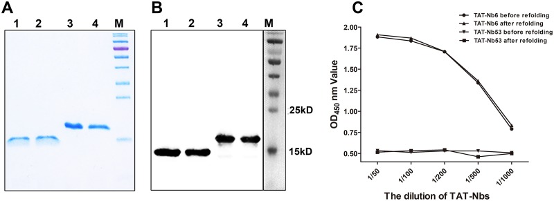
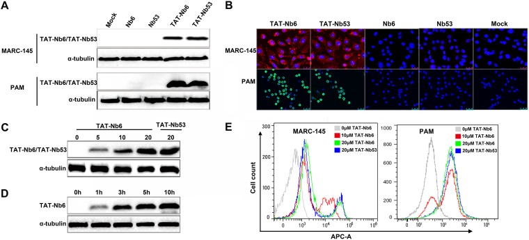
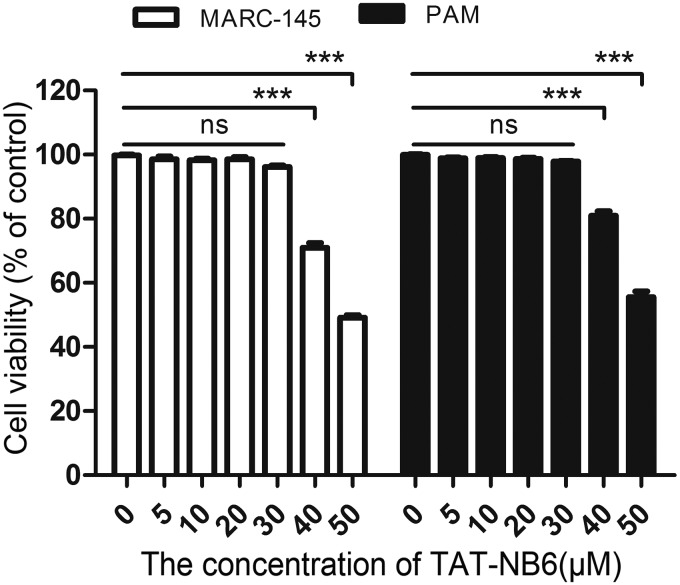
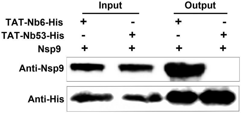
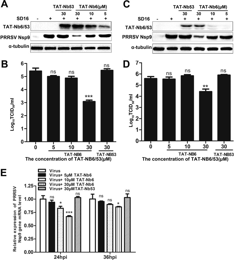
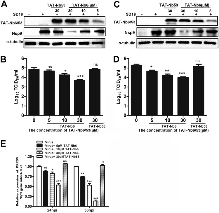
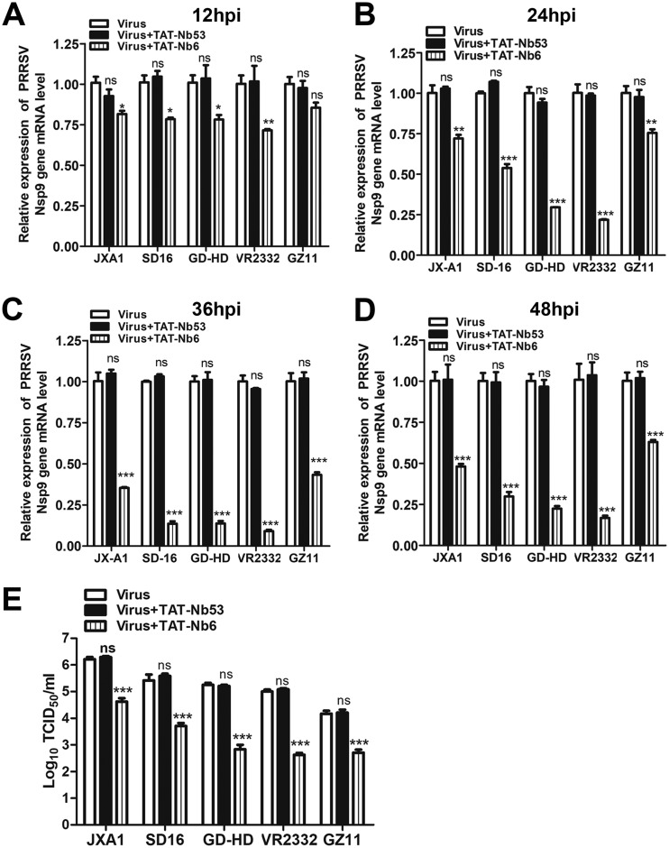
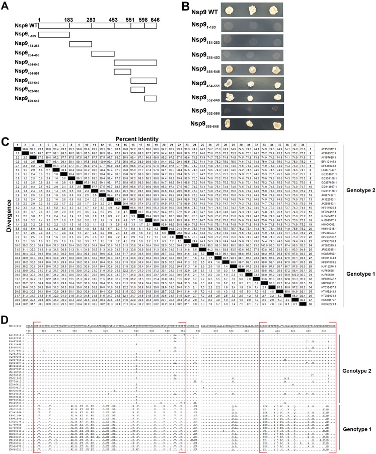
Porcine reproductive and respiratory syndrome (PRRS) is of great concern to the swine industry due to pandemic outbreaks of the disease, current ineffective vaccinations, and a lack of efficient antiviral strategies. In our previous study, a PRRSV Nsp9-specific nanobody, Nb6, was successfully isolated, and the intracellularly expressed Nb6 could dramatically inhibit PRRSV replication in MARC-145 cells. However, despite its small size, the application of Nb6 protein in infected cells is greatly limited, as the protein itself cannot enter the cells physically. In this study, a trans-activating transduction (TAT) peptide was fused with Nb6 to promote protein entry into cells. TAT-Nb6 was expressed as an inclusion body in Escherichia coli, and indirect enzyme-linked immunosorbent assays and pulldown assays showed that E. coli-expressed TAT-Nb6 maintained the binding ability to E. coli-expressed or PRRSV-encoded Nsp9. We demonstrated that TAT delivered Nb6 into MARC-145 cells and porcine alveolar macrophages (PAMs) in a dose- and time-dependent manner, and TAT-Nb6 efficiently inhibited the replication of several PRRSV genotype 2 strains as well as a genotype 1 strain. Using a yeast two-hybrid assay, Nb6 recognition sites were identified in the C-terminal part of Nsp9 and spanned two discontinuous regions (Nsp9aa454-551 and Nsp9aa599-646). Taken together, these results suggest that TAT-Nb6 can be developed as an antiviral drug for the inhibition of PRRSV replication and controlling PRRS disease.IMPORTANCE The pandemic outbreak of PRRS, which is caused by PRRSV, has greatly affected the swine industry. We still lack an efficient vaccine, and it is an immense challenge to control its infection. An intracellularly expressed Nsp9-specific nanobody, Nb6, has been shown to be able to inhibit PRRSV replication in MARC-145 cells. However, its application is limited, because Nb6 cannot physically enter cells. Here, we demonstrated that the cell-penetrating peptide TAT could deliver Nb6 into cultured cells. In addition, TAT-Nb6 fusion protein could suppress the replication of various PRRSV strains in MARC-145 cells and PAMs. These findings may provide a new approach for drug development to control PRRS.

Keywords: PRRSV; antiviral agents; nanobody.


Figures

FIG 1
FIG 1
Analysis of purified and refolded nanobodies by SDS-PAGE (A) and Western blotting (B). The predicted sizes of the His-tagged nanobodies are 15 kDa without TAT (NB6 and NB53) and 19 kDa with the TAT leader peptide (TAT-NB6 and TAT-NB53). M, protein marker; lanes 1 to 4, NB6, NB53, TAT-NB6, and TAT-NB53. (C) Determination of the binding activity of TAT-Nb6 to Nsp9 by iELISA. TAT-Nb53 was used as a negative control. Assays were performed in triplicate, and data are presented as means ± standard deviations (SD).
FIG 2
FIG 2
Cellular uptake of TAT-Nbs into MACR-145 and PAMs. (A and B) Western blotting (A) and IFA detection (B) of intracellular TAT-Nbs. MARC-145 and PAMs were treated with 10 μM Nb6, Nb53, TAT-Nb6, and TAT-Nb53 for 5 h. (C and E) Western blot (C) and flow cytometry (E) analyses of the uptake of TAT-NB6 and TAT-NB53 at different concentrations. MARC-145 cells were treated with the nanobodies at the indicated concentrations for 5 h. (D) MARC-145 cells were treated with TAT-NB6 at 20 μM for 0, 1, 3, 5, and 10 h and then examined by Western blotting.
FIG 3
FIG 3
Detection of TAT-Nb6 toxicity using CCK-8 kits. Data are expressed as means ± SD from three independent experiments. P values were calculated using ANOVA. P values were <0.05 (*), <0.01 (**), and <0.001 (***) compared with the cells infected with PRRSV alone (ns, not significant).
FIG 4
FIG 4
TAT-Nb6 interacts with PRRSV-encoded Nsp9. MARC-145 cells were infected with SD16 at an MOI of 1 for 48 h, and Nsp9 was pulled down by TAT-Nb6-His or TAT-Nb53-His. The bound proteins were detected by Western blotting using mouse anti-His antibody or mouse anti-Nsp9 antiserum.
FIG 5
FIG 5
Inhibition of PRRSV SD16 infection and replication by TAT-NB6 in MARC-145 cells. MARC-145 cells were infected with SD16 at an MOI of 0.01 for 1 h, and then the cell culture media were replaced with fresh DMEM containing 3% FBS and TAT-Nbs at the indicated concentrations. TAT-Nbs and PRRSV Nsp9 were detected at 24 hpi (A) and 36 hpi (C) by Western blotting using anti-His MAb and mouse anti-Nsp9 antiserum, respectively. Progeny virus released in the cell medium was measured by TCID50 at 24 hpi (B) and 36 hpi (D). (E) Relative levels of PRRSV RNA were detected by RT-qPCR using PRRSV Nsp9-specific primers. The GAPDH mRNA level served as an internal reference. Data are expressed as means ± SD from three independent experiments. P values were calculated using ANOVA as <0.05 (*), <0.01 (**), and <0.001 (***) compared with cells infected with PRRSV alone (ns, not significant).
FIG 6
FIG 6
Inhibition of PRRSV SD16 infection and replication by TAT-NB6 in PAMs. PAMs were infected with SD16 at an MOI of 0.01 for 1 h, and then the cell culture media were replaced with fresh RPMI 1640 containing 3% FBS and TAT-Nbs at the indicated concentrations. TAT-Nbs and PRRSV Nsp9 were detected at 24 hpi (A) and 36 hpi (C) by Western blotting using anti-His MAb and mouse anti-Nsp9 antiserum, respectively. Progeny virus released in the cell medium was measured by TCID50 at 24 hpi (B) and 36 hpi (D). (E) Relative levels of PRRSV RNA were detected by RT-qPCR using PRRSV Nsp9-specific primers. The GAPDH mRNA level served as an internal reference. Data are expressed as means ± SD from three independent experiments. P values were calculated using ANOVA as <0.05 (*), <0.01 (**), and <0.001 (***) compared with cells infected with PRRSV alone (ns, not significant).
FIG 7
FIG 7
TAT-NB6 inhibits the replication of multiple PRRSV strains. PAMs were infected with JXA1, SD16, GD-HD, VR2332, or GZ11-G1 at an MOI of 0.01 and incubated with 30 μM TAT-NB6 or TAT-NB53. The transcription levels of PRRSV Nsp9 in the cells were detected via RT-qPCR at 12 (A), 24 (B), 36 (C), and 48 (D) hpi. (E) The titers of progeny virus in the cell culture supernatants were measured by TCID50 at 48 hpi. Data are expressed as means ± SD from three independent experiments. P values were calculated using ANOVA as <0.05 (*), <0.01 (**), and <0.001 (***) compared with cells infected with PRRSV alone (ns, not significant).
FIG 8
FIG 8
Nb6 binds to the C-terminal end of Nsp9. (A) Nsp9 fragments used in this study for the identification of the Nb6-Nsp9 interaction domain. Full-length Nsp9 was truncated into four fragments in the first stage: Nsp1–183, Nsp9184–283, Nsp9284–453, and Nsp454–646. The C-terminal fragment, Nsp9454–646, was subsequently split into fragments Nsp9454–551, Nsp9552–646, Nsp9552–589, and Nsp9599–646. (B) Yeast two-hybrid assay. Vectors expressing the Nb6 protein and the indicated Nsp9 fragments were cotransformed into yeast cells. (C and D) Amino acid sequence comparison and alignment of Nsp9aa454–646. The sequences of Nsp9 from the 38 PRRSV strains in GenBank were analyzed using the Clustal W module of Lasergene 7.1 (DNASTAR, Inc.). Nb6-Nsp9 binding domains are shown in red square brackets, with dots indicating identical amino acid residues.

Similar articles

Cited by

References

    1. Liu H, Wang Y, Duan H, Zhang A, Liang C, Gao J, Zhang C, Huang B, Li Q, Li N, Xiao S, Zhou E-M. 2015. An intracellularly expressed Nsp9-specific nanobody in MARC-145 cells inhibits porcine reproductive and respiratory syndrome virus replication. Vet Microbiol 181:252–260. doi:10.1016/j.vetmic.2015.10.021. - DOI - PubMed
    1. Li N, Du T, Yan Y, Zhang A, Gao J, Hou G, Xiao S, Zhou EM. 2016. MicroRNA let-7f-5p inhibits porcine reproductive and respiratory syndrome virus by targeting MYH9. Sci Rep 6:34332. doi:10.1038/srep34332. - DOI - PMC - PubMed
    1. Zhang A, Zhao L, Li N, Duan H, Liu H, Pu F, Zhang G, Zhou EM, Xiao S. 2017. Carbon monoxide inhibits porcine reproductive and respiratory syndrome virus replication by the cyclic GMP/protein kinase G and NF-kappaB signaling pathway. J Virol 91:e01866-16. - PMC - PubMed
    1. Du Y, Du T, Shi Y, Zhang A, Zhang C, Diao Y, Jin G, Zhou E-M. 2016. Synthetic Toll-like receptor 7 ligand inhibits porcine reproductive and respiratory syndrome virus infection in primary porcine alveolar macrophages. Antiviral Res 131:9–18. doi:10.1016/j.antiviral.2016.04.005. - DOI - PubMed
    1. Goyal SM. 1993. Porcine reproductive and respiratory syndrome. J Vet Diagn Investig 5:656–664. doi:10.1177/104063879300500435. - DOI - PubMed

Publication types

MeSH terms

LinkOut - more resources