. 2019 Feb 5;93(4):e01764-18.
doi: 10.1128/JVI.01764-18. Print 2019 Feb 15.

The Host DHX9 DExH-Box Helicase Is Recruited to Chikungunya Virus Replication Complexes for Optimal Genomic RNA Translation

Affiliations

The Host DHX9 DExH-Box Helicase Is Recruited to Chikungunya Virus Replication Complexes for Optimal Genomic RNA Translation

Roy Matkovic et al. J Virol. .

Abstract

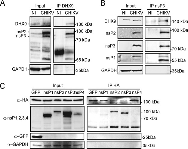
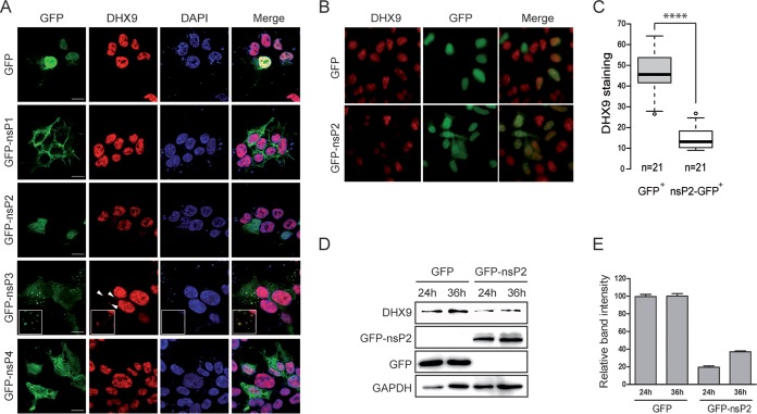
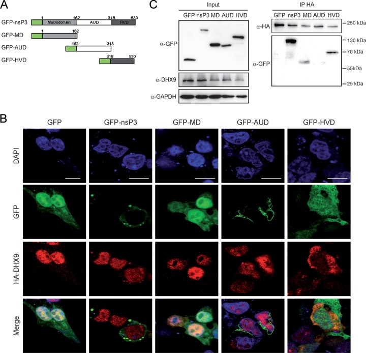
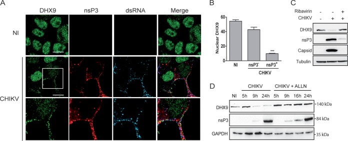
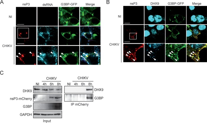
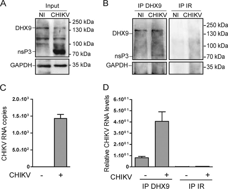
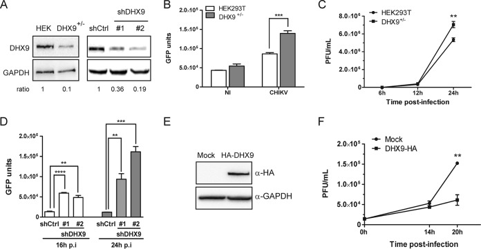
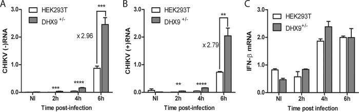
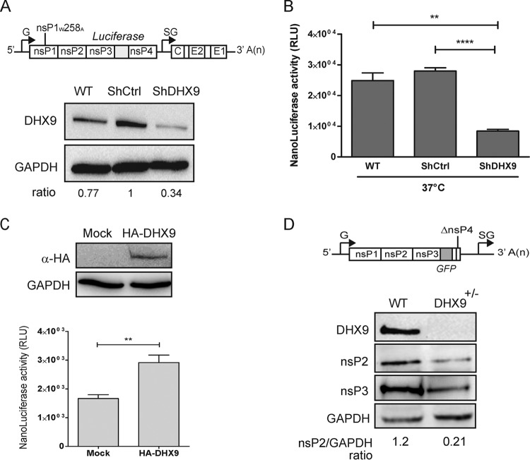
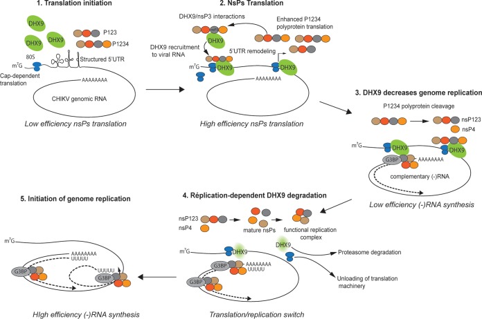
Beyond their role in cellular RNA metabolism, DExD/H-box RNA helicases are hijacked by various RNA viruses in order to assist replication of the viral genome. Here, we identify the DExH-box RNA helicase 9 (DHX9) as a binding partner of chikungunya virus (CHIKV) nsP3 mainly interacting with the C-terminal hypervariable domain. We show that during early CHIKV infection, DHX9 is recruited to the plasma membrane, where it associates with replication complexes. At a later stage of infection, DHX9 is, however, degraded through a proteasome-dependent mechanism. Using silencing experiments, we demonstrate that while DHX9 negatively controls viral RNA synthesis, it is also required for optimal mature nonstructural protein translation. Altogether, this study identifies DHX9 as a novel cofactor for CHIKV replication in human cells that differently regulates the various steps of CHIKV life cycle and may therefore mediate a switch in RNA usage from translation to replication during the earliest steps of CHIKV replication.IMPORTANCE The reemergence of chikungunya virus (CHIKV), an alphavirus that is transmitted to humans by Aedes mosquitoes, is a serious global health threat. In the absence of effective antiviral drugs, CHIKV infection has a significant impact on human health, with chronic arthritis being one of the most serious complications. The molecular understanding of host-virus interactions is a prerequisite to the development of targeted therapeutics capable to interrupt viral replication and transmission. Here, we identify the host cell DHX9 DExH-Box helicase as an essential cofactor for early CHIKV genome translation. We demonstrate that CHIKV nsP3 protein acts as a key factor for DHX9 recruitment to replication complexes. Finally, we establish that DHX9 behaves as a switch that regulates the progression of the viral cycle from translation to genome replication. This study might therefore have a significant impact on the development of antiviral strategies.

Keywords: DHX9; RNA helicase; chikungunya virus; nsP3; viral replication.


Figures

FIG 1
FIG 1
DHX9 helicase interacts with CHIKV nsPs in infected cells. Total lysates prepared from uninfected HeLa cells (NI) or from cells infected with CHIKV for 8 h were immunoprecipitated with anti-DHX9 (A) or anti-nsP3 serum (B). Samples of lysates (Input) and immunoprecipitates (IP) were separated by SDS-PAGE and probed with antisera against DHX9, CHIKV nsP1, nsP2, nsP3, or GAPDH MAbs as indicated. (C) HEK293T cells were cotransfected to express HA-DHX9, together with GFP or GFP-fused nsPs, as indicated. Cell lysates were prepared 36 h after transfection, precipitated with anti-HA agarose affinity beads, and separated by SDS-PAGE. Lysates and precipitated complexes were probed with MAbs against GFP, HA, or GAPDH. For each panel, the molecular masses are indicated.
FIG 2
FIG 2
GFP-nsP2 and GFP-nsP3 modulate DHX9 nuclear expression and subcellular localization, respectively. (A) HEK293T cells transfected to express either GFP or each of the GFP-fused nsPs (green) were stained for endogenous DHX9 (red) and analyzed by confocal microscopy. Nuclei were stained with DAPI (blue). Colocalized signals are indicated by arrows, and enlargements are shown in the inset. Scale bars, 5 μm. (B) Wide-field fluorescence microscopy of cells expressing the GFP-nsP2 protein or GFP alone and stained for endogenous DHX9. (C) Intensity of DHX9 nuclear staining was determined from cultures shown in panel B using ImageJ software. The number of cells analyzed for each condition is indicated. Mean values were compared using a Student t test. ****, P < 0.0001. (D) The endogenous DHX9 level in HEK293T cells was monitored by immunoblot analysis at various times after transfection of GFP or GFP-nsP2 expression plasmids. (E) The DHX9 band intensity in panel D was determined using ImageJ software. The results were normalized to the GAPDH signal and are expressed as percentages of the GFP condition.
FIG 3
FIG 3
CHIKV nsP3 binds DHX9 through its C-terminal HVD. (A) Schematic representation of GFP-nsP3 and its truncated derivatives. (B) HEK293T cells transfected to coexpress HA-DHX9, together with proteins depicted in panel A or with GFP, were stained with anti-DHX9 serum. Nuclei were stained with DAPI, and the cells were imaged by confocal microscopy. Scale bar, 5 μm. (C) Total cell extracts were prepared for each condition and precipitated with anti-HA agarose affinity beads. Proteins and bound complexes were visualized using antibodies against GFP, HA, or GAPDH, as indicated. Molecular masses are indicated.
FIG 4
FIG 4
Consequences of CHIKV infection on DHX9 levels and its subcellular localization. (A) HeLa cells infected for 8 h with the CHIKV-377-mCherry virus (MOI = 0.5) were fixed and stained with antibodies against DHX9 and dsRNA. Uninfected cells (NI) are shown as a control. Scale bars, 10 μm. (B) The intensity of DHX9 nuclear staining in nsP3+ or nsP3 cells from CHIKV infection condition or in uninfected (NI) culture was determined using ImageJ software. Mean values were compared using a Student t test. ***, P < 0.001. (C) Total lysate of uninfected cells or that from cells infected with CHIKV for 16 h in the absence or presence of ribavirin were probed with anti-DHX9, anti-nsP3, and anti-capsid antibodies; anti-tubulin MAb was used as a loading control. (D) Infected cells maintained in medium alone (CHIKV) or culture in the presence of 10 μM ALLN (CHIKV+ALLN) were collected at different times postinfection and analyzed by immunoblotting with antibodies specific to DHX9, nsP3, and GAPDH.
FIG 5
FIG 5
DHX9 is recruited to nsP3/G3BP complexes formed in the cytoplasm of CHIKV-infected cells. HeLa cells transfected to express the G3BP-GFP protein were left uninfected (NI) or were infected with the CHIKV-377-mCherry virus for 8 h. Cells were stained with anti-dsRNA antibodies (A) or anti-DHX9 serum (B) and processed for confocal imaging. Arrows indicate cytoplasmic foci with colocalized signals. For each panel, the lower lane shows an enlargement of the boxed zone. (C) Protein extracts prepared from uninfected cells or cells infected with the CHIKV-377-mCherry reporter virus for the indicated time were processed for immunoblot analysis of DHX9, nsP3, and G3PB expression (Input). Proteins complexed with nsP3 were immunoprecipitated (IP) with anti-mCherry MAbs and revealed by specific antibodies, as indicated.
FIG 6
FIG 6
DHX9/nsP3 complexes formed in CHIKV-infected cells contain viral RNA. (A) Cytosolic extracts prepared from uninfected cells (NI) or from cells infected for 6 h with CHIKV-LR-5'GFP (CHIKV) were analyzed for DHX9, nsP3, and GAPDH expression. (B) Complexes immunoprecipitated with anti-DHX9 rabbit polyclonal serum or nonspecific IgG (IR) were analyzed for the presence of DHX9, nsP3, and GAPDH using specific antibodies. (C and D) RNA isolated from cytosolic fractions (C) or from immunoprecipitates prepared using the same concentration of cytosolic proteins for each condition (D) was subjected to qRT-PCR with primers specific for CHIKV RNA. For input samples, values were normalized according to GAPDH mRNA copies. Values are means of duplicates.
FIG 7
FIG 7
Consequences of DHX9 expression on CHIKV infection. (A) Parental HEK293T cells, cells edited by CRISPR/Cas9 (DHX9+/–), or cells transfected with nontargeting shRNA (shCtrl) or shRNA against DHX9 (#1 and #2) were analyzed for DHX9 and GAPDH expression by Western blotting. The DHX9/GAPDH ratio determined by band intensity analysis is indicated for each condition. (B) Replication of the CHIKV-LR-5′GFP reporter virus in DHX9+/– cells and in the parental cell line was monitored by quantification of GFP expression in total cell extracts. Values are a mean of triplicate experiments. (C) Each cell line was infected with CHIKV-LR. Viral production in culture supernatants was determined by a standard plaque assay at the indicated times postinfection. Values are means of triplicates. (D) Replication of the CHIKV-LR-5′GFP reporter virus in cells transfected with shCtrl or shRNA against DHX9 was monitored by quantification of GFP expression in total cell extracts. Values are a mean of triplicate experiments. (E) Cells transfected with an empty plasmid (Mock) or with a plasmid encoding a HA-DHX9 protein were analyzed by immunoblotting with the indicated antibodies. (F) Cells were infected with CHIKV-LR, and virus production was monitored overtime in culture supernatant by a plaque assay. Mock-transfected cells are shown as a control. Values are means of triplicates; values were compared using a Student t test (*, P < 0.05; **, P < 0.01; ***, P < 0.001; ****, P < 0.0001).
FIG 8
FIG 8
DHX9 overexpression negatively regulates viral RNA synthesis independently of type I IFN expression. DHX9+/– cells and the parental HEK293T cells infected with the CHIKV-LR-5′GFP reporter virus for the indicated times were processed for qRT-PCR amplification of CHIKV (–)RNA (A) or (+)RNA (B). Noninfected cells (NI) are shown as a control. RNA levels were normalized according to GAPDH mRNA levels in the samples. Values were then normalized against an internal control, and results are expressed as arbitrary units. Analyses were performed in triplicate, and the error bars represent the standard deviations. (C) IFN-β mRNA was quantified from the same samples, as previously reported (74). GAPDH was used as an mRNA housekeeping control. Values are means of duplicates, and the error bars represent the standard deviations.
FIG 9
FIG 9
DHX9 expression regulates nonstructural protein translation. (A) Mock-transfected HEK293T cells (WT) and cells transfected with the indicated shRNAs were analyzed for DHX9 and GAPDH expression. The cells were infected with CHIKV-nsP1W258A-NanoLuc (depicted in the upper panel). (B) After 4 h at a nonpermissive temperature (37°C), the nanoluciferase activity was determined in cell lysates and normalized to the protein concentration in the samples. ****, P < 0.0001; **, P < 0.01. (C) HEK293T cells transfected to overexpress HA-DHX9 were infected CHIKV-nsP1W258A-NanoLuc and maintained at 37°C for 4 h. NsP translation was monitored by quantification of the nanoluciferase activity in the cell lysate, and values were normalized according to the protein content in the sample. The expression of the HA-DHX9 transgene was controlled by immunoblotting the total cell extracts with the indicated antibodies. (D) Parental (WT) or DHX9 CRISPR/Cas9-edited HEK293T cells were transfected with the ΔnsP4 CHIKV replicon (depicted in the upper panel). At 8 h after transfection, cell lysates were analyzed for DHX9, GAPDH, and nsP expression. The band intensity was determined, and the nsP2/GAPDH ratios are indicated.
FIG 10
FIG 10
Model of DHX9-dependent regulation of the alphavirus translation-to-replication switch. DHX9 is recruited to the plasma membrane-bound replication complexes through interactions with the HVD domain in nsP3, where it enhances nsP translation. As translation and nsP precursor cleavage proceeds, the concentration of mature nsP2 increases, accounting for DHX9 unloading from the replication complex and redirection to proteasomal degradation. In the absence of DHX9, translation is shut down, and replication starting with (–)RNA synthesis is favored. G3BP recruitment to replication complexes is depicted in this model.

Similar articles

Cited by

References

    1. Thiberville SD, Moyen N, Dupuis-Maguiraga L, Nougairede A, Gould EA, Roques P, de Lamballerie X. 2013. Chikungunya fever: epidemiology, clinical syndrome, pathogenesis, and therapy. Antiviral Res 99:345–370. doi:10.1016/j.antiviral.2013.06.009. - DOI - PMC - PubMed
    1. Rupp JC, Sokoloski KJ, Gebhart NN, Hardy RW. 2015. Alphavirus RNA synthesis and nonstructural protein functions. J Gen Virol 96:2483–2500. doi:10.1099/jgv.0.000249. - DOI - PMC - PubMed
    1. Spuul P, Balistreri G, Kaariainen L, Ahola T. 2010. Phosphatidylinositol 3-kinase-, actin-, and microtubule-dependent transport of Semliki Forest virus replication complexes from the plasma membrane to modified lysosomes. J Virol 84:7543–7557. doi:10.1128/JVI.00477-10. - DOI - PMC - PubMed
    1. Ahola T, Lampio A, Auvinen P, Kaariainen L. 1999. Semliki Forest virus mRNA capping enzyme requires association with anionic membrane phospholipids for activity. EMBO J 18:3164–3172. doi:10.1093/emboj/18.11.3164. - DOI - PMC - PubMed
    1. Lemm JA, Rumenapf T, Strauss EG, Strauss JH, Rice CM. 1994. Polypeptide requirements for assembly of functional Sindbis virus replication complexes: a model for the temporal regulation of minus- and plus-strand RNA synthesis. EMBO J 13:2925–2934. doi:10.1002/j.1460-2075.1994.tb06587.x. - DOI - PMC - PubMed

Publication types

MeSH terms