

Protein and lipid fingerprinting of native-like membrane complexes by combining TLC and protein electrophoresis
- PMID: 30463985
- PMCID: PMC6358299
- DOI: 10.1194/jlr.D090639
Protein and lipid fingerprinting of native-like membrane complexes by combining TLC and protein electrophoresis
Abstract
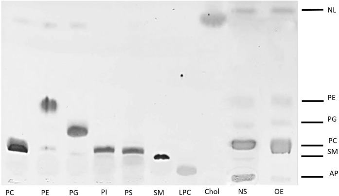
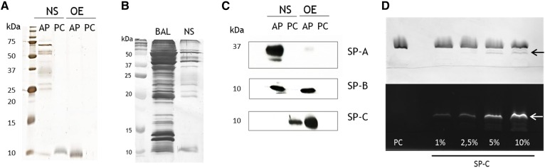
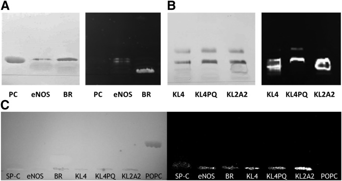
TLC has traditionally been used to analyze lipids isolated from membrane complexes. Here, we describe a method based on the combination of TLC and SDS-PAGE to qualitatively analyze the protein/lipid profile of membrane complexes such as those of lung surfactant. For this purpose, native lung surfactant was applied onto a silica TLC plate in the form of an aqueous suspension, preserving not only hydrophilic proteins associated with lipids but also native protein-lipid interactions. Using native membrane complexes in TLC allows the differential migration of lipids and their separation from the protein components. As a result, (partly) delipidated protein-enriched bands can be visualized and analyzed by SDS-PAGE to identify proteins originally associated with lipids. Interestingly, the hydrophobic surfactant protein C, which interacts tightly with lipids in native membrane complexes, migrates through the TLC plate, configuring specific bands that differ from those corresponding to lipids or proteins. This method therefore allows the detection and analysis of strong native-like protein-lipid interactions.
Keywords: diagnostic tools; lipoprotein; lung surfactant; phospholipids; proteomics; thin-layer chromatography.
Copyright © 2019 Lopez-Rodriguez et al.
Figures
Similar articles
-
High-resolution separation and quantification of neutral lipid and phospholipid species in mammalian cells and sera by multi-one-dimensional thin-layer chromatography.Anal Biochem. 1998 Apr 10;258(1):109-17. doi: 10.1006/abio.1997.2545. Anal Biochem. 1998. PMID: 9527856
-
The effect of lithium therapy upon the composition of the human erythrocyte membrane.J Inorg Biochem. 1995 Jan;57(1):23-32. doi: 10.1016/0162-0134(94)00010-8. J Inorg Biochem. 1995. PMID: 7876833
-
Coupling of native liquid phase isoelectrofocusing and blue native polyacrylamide gel electrophoresis: a potent tool for native membrane multiprotein complex separation.J Proteome Res. 2008 Mar;7(3):1326-40. doi: 10.1021/pr700613r. Epub 2008 Feb 5. J Proteome Res. 2008. PMID: 18247556
-
Solubilization of membrane protein complexes for blue native PAGE.J Proteomics. 2008 Aug 21;71(3):277-83. doi: 10.1016/j.jprot.2008.05.004. Epub 2008 Jun 6. J Proteomics. 2008. PMID: 18573355 Review.
-
Lipids in membrane protein structures.Biochim Biophys Acta. 2004 Nov 3;1666(1-2):2-18. doi: 10.1016/j.bbamem.2004.06.012. Biochim Biophys Acta. 2004. PMID: 15519305 Review.
Cited by
-
Application of lipidomics in the study of traditional Chinese medicine.J Pharm Anal. 2025 Feb;15(2):101083. doi: 10.1016/j.jpha.2024.101083. Epub 2024 Aug 30. J Pharm Anal. 2025. PMID: 39995576 Free PMC article. Review.
-
Egg yolk lipids: separation, characterization, and utilization.Food Sci Biotechnol. 2022 Aug 10;31(10):1243-1256. doi: 10.1007/s10068-022-01138-4. eCollection 2022 Sep. Food Sci Biotechnol. 2022. PMID: 35992319 Free PMC article. Review.
References
-
- Skipski V. P., Barclay M., Reichman E. S., and Good J. J.. 1967. Separation of acidic phospholipids by one-dimensional thin-layer chromatography. Biochim. Biophys. Acta. 137: 80–89. - PubMed
-
- Skipski V. P., Peterson R. F., Sanders J., and Barclay M.. 1963. Thin-layer chromatography of phospholipids using silica gel without calcium sulfate binder. J. Lipid Res. 4: 227–228. - PubMed
-
- Allan D., and Cockcroft S.. 1982. A modified procedure for thin-layer chromatography of phospholipids. J. Lipid Res. 23: 1373–1374. - PubMed
-
- Weerheim A. M., Kolb A. M., Sturk A., and Nieuwland R.. 2002. Phospholipid composition of cell-derived microparticles determined by one-dimensional high-performance thin-layer chromatography. Anal. Biochem. 302: 191–198. - PubMed
Publication types
MeSH terms
Substances
LinkOut - more resources
Full Text Sources
Research Materials
