. 2019 Feb 14;133(7):697-709.
doi: 10.1182/blood-2018-10-881722. Epub 2018 Nov 21.

GM-CSF inhibition reduces cytokine release syndrome and neuroinflammation but enhances CAR-T cell function in xenografts

Affiliations

GM-CSF inhibition reduces cytokine release syndrome and neuroinflammation but enhances CAR-T cell function in xenografts

Rosalie M Sterner et al. Blood. .

Abstract

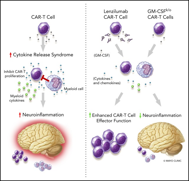
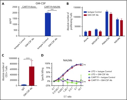
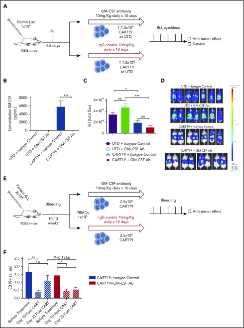
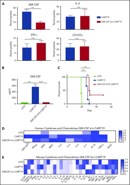
Chimeric antigen receptor T (CAR-T) cell therapy is a new pillar in cancer therapeutics; however, its application is limited by the associated toxicities. These include cytokine release syndrome (CRS) and neurotoxicity. Although the IL-6R antagonist tocilizumab is approved for treatment of CRS, there is no approved treatment of neurotoxicity associated with CD19-targeted CAR-T (CART19) cell therapy. Recent data suggest that monocytes and macrophages contribute to the development of CRS and neurotoxicity after CAR-T cell therapy. Therefore, we investigated neutralizing granulocyte-macrophage colony-stimulating factor (GM-CSF) as a potential strategy to manage CART19 cell-associated toxicities. In this study, we show that GM-CSF neutralization with lenzilumab does not inhibit CART19 cell function in vitro or in vivo. Moreover, CART19 cell proliferation was enhanced and durable control of leukemic disease was maintained better in patient-derived xenografts after GM-CSF neutralization with lenzilumab. In a patient acute lymphoblastic leukemia xenograft model of CRS and neuroinflammation (NI), GM-CSF neutralization resulted in a reduction of myeloid and T cell infiltration in the central nervous system and a significant reduction in NI and prevention of CRS. Finally, we generated GM-CSF-deficient CART19 cells through CRISPR/Cas9 disruption of GM-CSF during CAR-T cell manufacturing. These GM-CSFk/o CAR-T cells maintained normal functions and had enhanced antitumor activity in vivo, as well as improved overall survival, compared with CART19 cells. Together, these studies illuminate a novel approach to abrogate NI and CRS through GM-CSF neutralization, which may potentially enhance CAR-T cell function. Phase 2 studies with lenzilumab in combination with CART19 cell therapy are planned.


Conflict of interest statement

Conflict-of-interest disclosure: S.S.K. is an inventor on patents in the field of CAR-T cell therapy that are licensed to Novartis (under an agreement among the Mayo Clinic, the University of Pennsylvania, and Novartis). O.A., T.S., D.C., and C.D. are employed by Humanigen. R.M.S., R.S., M.J.C., and S.S.K. are inventors on patents related to this work. The remaining authors declare no competing financial interests.

Figures

None
Graphical abstract
Figure 1.
Figure 1.
GM-CSF neutralization in vitro enhances CAR-T cell proliferation in the presence of monocytes and does not impair CAR-T–cell effector function. (A) Lenzilumab neutralizes CAR-T cell–produced GM-CSF in vitro compared with isotype-control treatment, as assayed by a multiplex assay after 3 days of culture with CART19 cells in media alone or CART19 cells cocultured with NALM6; n = 2 experiments, 2 replicates per experiment, a representative experiment is depicted. (B) GM-CSF–neutralizing antibody treatment did not inhibit the ability of CAR-T cells to proliferate, as assayed by a carboxyfluorescein diacetate succinimidyl ester flow cytometry proliferation assay of live CD3 cells; n = 3 donors, 2 replicates per donor, a representative experiment at the 3-day time point is depicted. (C) Lenzilumab enhanced the proliferation of CART19 cells compared with isotype-control–treated with CART19 cells when cocultured with monocytes; n = 3 donors at the 3-day time point, 2 replicates per donor. (D) Lenzilumab treatment did not inhibit cytotoxicity of CART19 cells or UTD T cells when cultured with NALM6; n = 3 donors, 2 replicates per donor, a representative experiment at the 48-hour time point is depicted. All data are mean ± SEM. ***P < .001, ****P < .0001, Student t test. Alone, CART19 cells in media alone; MOLM13, CART19+MOLM13; ns, P > .05, lenzilumab vs isotype-control treatment using the Student t test; NALM6, CART19+NALM6; PMA/ION, CART19 cells plus 5 ng/mL PMA and 0.1 μg/mL ionomycin.
Figure 2.
Figure 2.
GM-CSF neutralization in vivo enhances CAR-T cell antitumor activity in xenograft models. (A) Experimental schema: NSG mice were injected IV with the luciferase+CD19+ cell line NALM6 (1 × 10⁶ cells per mouse). Four to six days later, mice were imaged and randomized, and they received 1 to 1.5 × 10⁶ CART19 cells or an equivalent number of total cells of control UTD cells the following day, with lenzilumab or control IgG (10 mg/kg, given intraperitoneally daily for 10 days, starting on the day of CAR-T injection). Mice were followed with serial bioluminescence imaging to assess disease burden beginning at day 7 post–CAR-T cell injection and were followed for overall survival. Tail vein bleeding was performed 7 or 8 days after CAR-T cell injection. (B) Lenzilumab neutralizes CAR-T–produced serum GM-CSF in vivo compared with isotype-control treatment, as assayed by a GM-CSF singleplex assay; n = 2 experiments, 7 or 8 mice per group, representative experiment, serum from day 8 post–CAR-T cell/UTD cell injection. Data are mean ± SEM. (C) Lenzilumab-treated CAR-T cells and isotype-control–treated CAR-T cells are equally effective at controlling tumor burden in vivo in a high tumor burden relapse xenograft model of ALL, day 7 post–CAR-T injection; n = 2 experiments, 7 or 8 mice per group, a representative experiment is depicted. Data are mean ± SEM. (D) Mouse images from (C). (E) Experimental schema: NSG mice were injected IV with the blasts derived from patients with ALL (1 × 10⁶ cells per mouse). Mice were bled serially and when the CD19+ cells were ∼1 per microliter, mice were randomized to receive 2.5 × 10⁶ CART19 cells with lenzilumab or control IgG (10 mg/kg, given intraperitoneally daily for 10 days, starting on the day of CAR-T injection). Mice were followed with serial tail vein bleeding to assess disease burden beginning at day 14 post–CAR-T cell injection and were followed for overall survival. (F) Lenzilumab treatment with CAR-T therapy results in more sustained control of tumor burden over time in a patient ALL xenograft model compared with isotype-control treatment with CAR-T therapy; 6 mice per group. Data are mean ± SEM. *P < .05, **P < .01, ***P < .001, Student t test. ns, P > .05, Student t test.
Figure 3.
Figure 3.
GM-CSF CRISPR-knockout CAR-T cells exhibit reduced expression of GM-CSF, similar levels of key cytokines and chemokines, and enhanced antitumor activity. (A) CRISPR Cas9 GM-CSFk/o CART19 cells exhibit reduced GM-CSF production compared with wild-type CART19 cells, but other cytokine production and degranulation are not inhibited by the GM-CSF gene disruption. CART19 cells and GM-CSFk/o CART19 cells stimulated with NALM6; n = 3 experiments, 2 replicates per experiment. Data are mean ± SEM. ***P < .001, Student t test. n.s., P > .05, Student t test. (B) GM-CSF k/o CAR-T cells have reduced serum human GM-CSF in vivo compared with CAR-T treatment, as assayed by multiplex; 5 or 6 mice per group (4 to 6 at time of bleed, 8 days post–CAR-T cell injection). Data are mean ± SEM. ***P < .001, ****P < .0001, Student t test. (C) GM-CSFk/o CART19 cells in vivo enhance overall survival compared with wild-type CART19 cells in a high tumor burden relapse xenograft model of ALL utilizing a NALM6 cell line; 5 or 6 mice per group. **P < .01, log-rank test. Human (D) and mouse (E) multiplex of serum cytokines and chemokines. A statistically significant reduction of human GM-CSF in GM-CSFk/o CART19 cells compared to wild-type CAR-T cells was observed, but no other statistically significant differences between GM-CSFk/o CART19 cells and wild-type CART19 cells were seen, further implicating that critical T-cell cytokines and chemokines are not adversely depleted by reducing GM-CSF expression; 5 or 6 mice per group (4 to 6 at time of bleed). ****P < .0001, Student t test.
Figure 4.
Figure 4.
Patient-derived xenograft model for NI and CRS. (A) Experimental schema: mice received 1 to 3 × 10⁶ blasts derived from the peripheral blood of patients with ALL. Mice were monitored for engraftment for ∼10 to 13 weeks via tail vein bleeding. When serum CD19+ cells were ≥10 per microliter, the mice received CART19 cells (2 to 5 × 10⁶ cells) and commenced antibody therapy for a total of 10 days, as indicated. Mice were weighed on a daily basis as a measure of their well-being. Mouse brain MRIs were performed 5 or 6 days post–CART19 cell injection, and tail vein bleeding for cytokine/chemokine and T cell analysis was performed 4 to 11 days post–CART19 cell injection; 2 independent experiments. (B) Combination of GM-CSF neutralization with CART19 cells is equally effective as isotype control antibodies combined with CART19 cells in controlling CD19+ burden of ALL cells; representative experiment, 3 mice per group, 11 days post–CART19 cell injection. Data are mean ± SEM. (C) Brain MRI with CART19 cell therapy exhibits T1 enhancement, suggestive of blood–brain barrier disruption and possible edema; 3 mice per group, 5 or 6 days post–CART19 cell injection, representative image. (D) High tumor burden ALL patient–derived xenografts (PDX) treated with CART19 cells show human CD3 cell infiltration of the brain compared with untreated PDX controls; 3 mice per group, representative image. *P < .05, Student t test.
Figure 5.
Figure 5.
Canonical pathways are altered in brains from patient-derived xenografts after treatment with CART19 cells. Red boxes indicate upregulation of genes in CART19 cells plus isotype control–treated mice compared with the untreated patient-derived xenografts.
Figure 6.
Figure 6.
GM-CSF neutralization in vivo ameliorates CRS after CART19 therapy in a xenograft model. (A) Lenzilumab and anti-mouse GM-CSF antibody prevent CRS-induced weight loss compared with mice treated with CART19 cells and isotype-control antibodies; 3 mice per group, 2-way analysis of variance. Data are mean ± SEM. (B) Human GM-CSF was neutralized in patient-derived xenografts treated with lenzilumab and mouse GM-CSF–neutralizing antibody; 3 mice per group. Data are mean ± SEM. *P < .05, ***P < .001, Student t test. (C) Human cytokine/chemokine heat map (serum collected 11 days after CART19 cell injection) exhibits increases in cytokines and chemokines typical of CRS after CART19 cell treatment. GM-CSF neutralization results in a significant decrease in several cytokines and chemokines compared with mice treated with CART19 cells and isotype-control antibodies, including several myeloid-associated cytokines and chemokines, as indicated in the panel; 3 mice per group, serum from day 11 post–CART19 cell injection. *P < .05, **P < .01, ***P < .001, GM-CSF neutralizing antibody–treated mice vs isotype control–treated mice that received CAR-T cell therapy, Student t test. (D) Mouse cytokine/chemokine heat map (serum collected 11 days after CART19 cell injection) exhibits increases in mouse cytokines and chemokines typical of CRS after CART19 cell treatment. GM-CSF neutralization results in a significant decrease in several cytokines and chemokines compared with treatment with CART19 cells with control antibodies, including several myeloid-differentiating cytokines and chemokines, as indicated in the panel; 3 mice per group, serum from day 11 post–CART19 cell injection, *P < .05, GM-CSF neutralizing antibody–treated mice vs isotype control–treated mice that received CAR-T cell therapy, Student t test.
Figure 7.
Figure 7.
GM-CSF neutralization in vivo ameliorates NI after CART19 cell therapy in a xenograft model. (A-B) Gadolinium-enhanced T1-hyperintensity (cubic millimeters) MRI showed that GM-CSF neutralization helped to reduced brain inflammation, blood–brain barrier disruption, and possible edema compared with isotype control. (A) Representative images. (B) Three mice per group. Data are mean ± SD. *P < .05, **P < .01, 1-way analysis of variance. (C) Human CD3 T cells were present in the brain after treatment with CART19 cell therapy. GM-CSF neutralization resulted in a decreased raw average of CD3 infiltration in the brain (although not statistically significant), as assayed by flow cytometry in brain hemispheres; 3 mice per group. Data are mean ± SEM. *P < .05, Student t test. (D) CD11b+ bright macrophages were decreased in raw average (although not statistically significant) in the brains of mice receiving GM-CSF neutralization during CAR-T cell therapy compared with those receiving isotype-control treatment during CAR-T cell therapy, as assayed by flow cytometry in brain hemispheres; 3 mice per group. Data are mean ± SEM, Student t test.

References

    1. Kenderian SS, Ruella M, Gill S, Kalos M. Chimeric antigen receptor T-cell therapy to target hematologic malignancies. Cancer Res. 2014;74(22):6383-6389. - PubMed
    1. Lim WA, June CH. The principles of engineering immune cells to treat cancer. Cell. 2017;168(4):724-740. - PMC - PubMed
    1. Neelapu SS, Locke FL, Bartlett NL, et al. . Axicabtagene ciloleucel CAR T-cell therapy in refractory large B-cell lymphoma. N Engl J Med. 2017;377(26):2531-2544. - PMC - PubMed
    1. Maude SL, Laetsch TW, Buechner J, et al. . Tisagenlecleucel in children and Young adults with B-cell lymphoblastic leukemia. N Engl J Med. 2018;378(5):439-448. - PMC - PubMed
    1. Schuster SJ, Svoboda J, Chong EA, et al. . Chimeric antigen receptor T cells in refractory B-cell lymphomas. N Engl J Med. 2017;377(26):2545-2554. - PMC - PubMed

Publication types

MeSH terms

Substances