Review
. 2018 Dec 7;62(6):829-838.
doi: 10.1042/EBC20180029. Print 2018 Dec 7.

Coordination of eukaryotic cilia and flagella

Affiliations
Review

Coordination of eukaryotic cilia and flagella

Kirsty Y Wan. Essays Biochem. .

Abstract

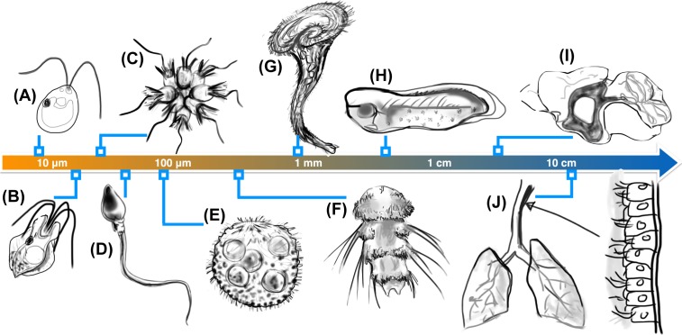
Propulsion by slender cellular appendages called cilia and flagella is an ancient means of locomotion. Unicellular organisms evolved myriad strategies to propel themselves in fluid environments, often involving significant differences in flagella number, localisation and modes of actuation. Remarkably, these appendages are highly conserved, occurring in many complex organisms such as humans, where they may be found generating physiological flows when attached to surfaces (e.g. airway epithelial cilia), or else conferring motility to male gametes (e.g. undulations of sperm flagella). Where multiple cilia arise, their movements are often observed to be highly coordinated. Here I review the two main mechanisms for motile cilia coordination, namely, intracellular and hydrodynamic, and discuss their relative importance in different ciliary systems.

Keywords: algae; centrioles/basal bodies; cilia; coordination; hydrodynamics; swimming.


Conflict of interest statement

The author declares that there are no competing interests associated with the manuscript.

Figures

Figure 1
Figure 1. Unity and diversity of ciliary systems
The same fundamental structure occurs in the tiniest of microorganisms as well as ciliated tissues, but exhibits drastic differences in number and localisation. Examples include (A) the algal biflagellate Chlamydomonas reinhardtii [4], (B) a quadriflagellate Prasinophyte alga Pyramimonas sp. [12], (C) rosette-forming choanoflagellates [13,14], (D) human sperm [15], (E) the spherical alga Volvox carteri [16,17], (F) the ciliated larvae of the marine annelid Platynereis dumerilii which have segmental multiciliated cells, and long, stiff chaetae [18], (G) the trumpet-shaped ciliate Stentor coeruleus [19], (H) ciliated epithelia of Xenopus laevis embryos [20], (I) ependymal cilia in mouse brain ventricles which direct cerebrospinal fluid flows [8] (cilia are localised to shaded region) and (J) multiciliated columnar cells in the human trachea [10,21].
Figure 2
Figure 2. The Chlamydomonas flagellar apparatus
(A) Schematic showing the cytoskeletal architecture of basal bodies (B1,2), microtubular roots (two-membered rootlets R2 and four-membered rootlets R4), and fibrous/contractile connections (NBBCs to the nucleus, proximal and distal striated fibres PF and DF). (B) Longitudinal section of a flagellum showing a structural change from triplet microtubules to doublets, two characteristic cross-sections are shown: one through the basal body and the second through flagellum proper. (C) Top view, highlighting radial symmetries in the flagellar apparatus, the locations of the two PB, the cruciate arrangement of microtubule bundles and the DF connecting specific microtubule doublets in the mature basal bodies (B1,2),
Figure 3
Figure 3. Experimental versus natural configurations of algal flagella
In all cases, the conspicuous orange dots represent algal eyespots (rudimentary photoreceptors), whose positioning is related to the developmental age of the flagella. A pair of V. carteri somatic cells held on nearby micropipettes, interacting hydrodynamically, exhibits either in-phase synchrony (A) or anti-phase synchrony (B) depending on their relative orientation. (C) In vivo, arrays of these cells coordinate metachronal waves in the Volvox colony. By contrast, the in-phase breaststroke of C. reinhardtii (D) cannot be reproduced in pairs of wildtype cells that have been rendered uniflagellate (E), implicating an internal (possibly spring-like) coupling provided by the distal striated fibre. Arrows indicate power stroke directions (A–E). (F) In a different species (see also Figure 1B), a quadriflagellate beat pattern (aka trot) is observed, comprising two pairs of breaststrokes displaced temporally by 1/2 beat cycle.
Figure 4
Figure 4. Aberrant flagellar coordination patterns in a basal-coupling mutant.
Flagellar waveforms and dynamics are tracked in a tri-flagellate vfl-3 mutant (A) and wildtype C. reinhardtii (B). (Cell body/pipette not shown on subsequent image frames.) The distal striated fibre is missing or defective in vfl-3 [81]. Consequently, coordination between the three flagella reverts to hydrodynamic interactions, in which the pair beating with power strokes in the same direction tends to synchronize in in-phase, but in anti-phase with respect to the third singlet flagellum. The wildtype cell on the other hand, maintains an in-phase synchronous breaststroke. (For further examples, see [12].)
Figure 5
Figure 5. Revealing intrinsic differences between the two C.reinhardtii flagella
The two flagella are similar but not identical. In vivo micromanipulation and microsurgery revealed a differential sensitivity to deflagellation-induced intracellular calcium elevation – here inferred indirectly by measuring ciliary beat frequencies in the two flagella of the same cell, after successive rounds of deflagellation and regrowth. The stereotypical trans-flagellum beating mode is markedly faster, has an attenuated waveform, and exhibits greater frequency variations than the cis.

References

    1. Darwin C.R. (1875) The Movements and Habits of Climbing Plants, John Murray, London
    1. Mitchell D.R. (2007) The evolution of eukaryotic cilia and flagella as motile and sensory organelles. Adv. Exp. Med. Biol. 607, 130–140 10.1007/978-0-387-74021-8_11 - DOI - PMC - PubMed
    1. Ishimoto K., Gadelha H., Gaffney E.A., Smith D.J. and Kirkman-Brown J. (2017) Coarse-graining the fluid flow around a human sperm. Phys. Rev. Lett. 118, 124501 10.1103/PhysRevLett.118.124501 - DOI - PubMed
    1. Ruffer U. and Nultsch W. (1987) Comparison of the beating of cis-flagella and trans-flagella of Chlamydomonas cells held on micropipettes. Cell Motil. Cytoskeleton 7, 87–93 10.1002/cm.970070111 - DOI - PubMed
    1. Goldstein R.E., Polin M. and Tuval I. (2009) Noise and synchronization in pairs of beating eukaryotic flagella. Phys. Rev. Lett. 103, 168103 10.1103/PhysRevLett.103.168103 - DOI - PubMed

Publication types