. 2018 Nov;563(7732):559-563.
doi: 10.1038/s41586-018-0702-1. Epub 2018 Nov 21.

Protocadherin-1 is essential for cell entry by New World hantaviruses

Affiliations

Protocadherin-1 is essential for cell entry by New World hantaviruses

Rohit K Jangra et al. Nature. 2018 Nov.

Abstract

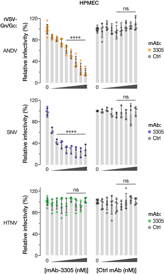
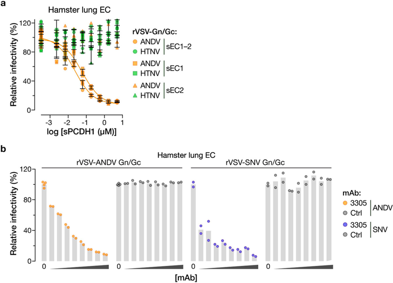
The zoonotic transmission of hantaviruses from their rodent hosts to humans in North and South America is associated with a severe and frequently fatal respiratory disease, hantavirus pulmonary syndrome (HPS)1,2. No specific antiviral treatments for HPS are available, and no molecular determinants of in vivo susceptibility to hantavirus infection and HPS are known. Here we identify the human asthma-associated gene protocadherin-1 (PCDH1)3-6 as an essential determinant of entry and infection in pulmonary endothelial cells by two hantaviruses that cause HPS, Andes virus (ANDV) and Sin Nombre virus (SNV). In vitro, we show that the surface glycoproteins of ANDV and SNV directly recognize the outermost extracellular repeat domain of PCDH1-a member of the cadherin superfamily7,8-to exploit PCDH1 for entry. In vivo, genetic ablation of PCDH1 renders Syrian golden hamsters highly resistant to a usually lethal ANDV challenge. Targeting PCDH1 could provide strategies to reduce infection and disease caused by New World hantaviruses.


Figures

Extended Data Fig. 1 |
Extended Data Fig. 1 |. PCDH1 is required for entry of New World hantaviruses into HAP1 and U2OS cells.
a, Genes enriched for genetrap insertions in the rVSV-ANDV Gn/Gc-selected population versus unselected control cells. The size of each bubble reflects the number of independent gene-trap insertions observed. Candidate genes are associated with cholesterol metabolism (yellow), the endoplasmic reticulum membrane complex network (green), and transcription (blue). PCDH1 (red) is a singleton hit. b, Single-cell HAP1 clones deficient for PCDH1 were generated by CRISPR–Cas9 genome engineering. The sequences of PCDH1-knockout alleles in clones 1 and 22 are shown. The sgRNA target sequence is highlighted in red. i1, insertion of a single nucleotide in PCDH1. c, WT and HAP1 PCDH-KO cell lines were exposed to rVSVs bearing the indicated viral glycoproteins at a MOI of 0.02 IU per cell. ‘+cDNA’ indicates complementation of PCDH1-KO cells with PCDH1. Cells were scored for infection at 20 hpi. One hundred per cent relative infection corresponds to 20–30% infected cells. Averages ± s.d. are shown: HAP1-WT, three experiments, n = 6; PCDH1-KO#1-cDNA, two experiments, n = 5; PCDH1-KO#1+cDNA, three experiments, n = 8; PCDH1-KO#22-cDNA, three experiments, n = 8, except for HTNV Gn/Gc, for which two experiments, n = 4; PCDH1- KO#1+cDNA, three experiments, n = 8; PCDH1-KO#22+cDNA, three experiments, n = 8. d, Single-cell U2OS clones deficient for PCDH1 were generated by CRISPR–Cas9 genome engineering. The sequences of PCDH1-knockout alleles in clone 5 are shown. The sgRNA target sequence is highlighted in red. i1, insertion of a single nucleotide in PCDH1. e,f, WT and U2OS PCDH1-KO cell lines were exposed to the indicated rVSVs at a MOI of 0.02 IU per cell. Cells were scored for infection at 20 hpi. eGFP-positive infected cells (pseudocoloured green) were detected by fluorescence microscopy (e) and enumerated by automated counting (f). One hundred per cent relative infection corresponds to 10–20% infected cells. Averages ± s.e.m. are shown, three experiments, n = 13. In c, f, wild type versus PCDH1-KO by two-way ANOVA with Tukey’s (c) or Sidak’s (f) tests: NS, P > 0.05; ****, P < 0.0001.
Extended Data Fig. 2 |
Extended Data Fig. 2 |. Expression and plasma-membrane localization of PCDH1 variants lacking domains EC1 or EC2 of PCDH1.
a, U2OS PCDH1-KO cell lines complemented with the indicated PCDH1 proteins were immunostained with an anti-Flag antibody and visualized by fluorescence microscopy. EV, empty vector. Scale bar, 20 μm. b, c, Live cells from a were stained with the PCDHl-EC7-specific mAb 3677 at 4 °C to detect cell-surface PCDH1 and visualized by flow cytometry. Cells were gated on PCDH1 immunofluorescence intensity (dotted red lines in c) to determine the percentage of cells with surface expression of each PCDH1 protein. Averages ± s.d. are shown in b: two experiments, n = 4, except in the case of WT, for which n = 3. c, Representative flow plots from b. Experiments were performed three times with similar results.
Extended Data Fig. 3 |
Extended Data Fig. 3 |. Expression, purification and characterization of soluble PCDH1 proteins.
a, Purified sEC1–2 bearing C-terminal Flag and decahistidine (His10) epitope tags was resolved on an SDS–polyacrylamide gel and visualized by Coomassie blue staining. Asterisk, minor component of unknown origin. Mr, relative molecular weight (K denotes × 1,000); M, monomer peak. The experiment was performed three times with similar results. b, SEC-MALS analysis of purified sEC1–2 from a. Absorbance (arbitrary units, au) was monitored at 280 nm; m, monomer peak. Calculated (MWcalc) and observed (MWobs) molecular-weight estimates from MALS are shown in the inset. The experiment was performed twice with similar results. c, Generation of purified sEC1, comprising the first extracellular cadherin domain of PCDH1, and bearing a C-terminal His10 epitope tag. sEC1–2 is shown for comparison. The experiment was performed three times with similar results. d, Capacity of sEC1 and sEC2 to block hantavirus glycoprotein-dependent entry. rVSVs bearing ANDV Gn/Gc were preincubated with sEC1 or sEC2 (0–5 μM, in serial threefold dilutions), and then allowed to infect HUVECs at a MOI of 0.2 IU per cell. Cells were scored for infection at 9 hpi. One hundred per cent relative infection corresponds to 10–20% infected cells. Averages ± s.d.: three experiments, n = 8 for sEC1; n = 7 for sEC2.
Extended Data Fig. 4 |
Extended Data Fig. 4 |. Isolation of PCDHl-specific monoclonal antibodies.
a, Flow chart showing isolation of PCDH1-specific mAbs from Library F by phage display. b, Capacity of selected mAb clones to recognize recombinant fusion proteins comprising PCDH1 ectodomains (containing or lacking EC1) and the Fc antibody domain. BSA, bovine serum albumin. Data from a representative ELISA experiment are shown. Experiments were performed three times with similar results. c, Kinetic binding analysis of selected Fabs to PCDH1(ectodomain)–Fc fusion proteins by surface plasmon resonance. nd, not determined; Kd, equilibrium dissociation constant. d, mAb 3305 recognizes the first extracellular cadherin (EC1) domain of PCDH1. U2OS PCDH1-KO cells, uncomplemented (KO) or complemented either with full-length PCDH1 (WT) or with a PCDH1 variant lacking the EC1 domain (ΔEC1), were co-immunostained with anti-Flag (α-Flag) mAb and anti-PCDH1 mAb 3305, or with anti-Flag and negative control antibodies (hIgG). Cells were visualized by fluorescence microscopy. Scale bar, 20 μm. Data from a representative experiment are shown. Experiments were performed three times with similar results.
Extended Data Fig. 5 |
Extended Data Fig. 5 |. PCDH1 ECl-specific mAb 3305 blocks ANDV and SNV entry into primary HPMECs.
HPMECs were preincubated with mAb 3305 or with human IgG control (Ctrl) (0–100 μg ml–1, 0–680 nM, in serial threefold dilutions), and then exposed to the indicated rVSVs Gn/Gc glycoproteins at a MOI of 0.2 IU per cell. Infected cells were scored at 9 hpi. One hundred per cent relative infectivity corresponds to 15–20% infected cells. Averages ± s.d.: three experiments; no-mAb samples (‘0’), n = 10 for ANDV and HTNV, n = 3 for SNV; mAb-treated samples, n = 5 for ANDV and HTNV, n = 3 for SNV. mAb 3305 versus control mAb, two-way ANO VA with Dunnett’s test: ns, P > 0.05; ****P < 0.0001.
Extended Data Fig. 6 |
Extended Data Fig. 6 |. The monoclonal antibody 1E11/D3 recognizes HTNV and ANDV Gn/Gc.
293FT cells transfected with plasmids encoding ANDV or HTNV Gn/Gc were analysed by flow cytometry using 1E11/D3 mAb for total (after permeabilization) and surface (without permeabilization) expression of Gn/Gc. Emptyvector-transfected cells were used as a negative control for gating. Results are from a representative experiment with n = 2. Experiments were performed five times for ANDV and twice for HTNV with similar results.
Extended Data Fig. 7 |
Extended Data Fig. 7 |. PCDH1 mediates viral entry by direct binding to hantavirus glycoproteins.
Capacity of hantavirus glycoproteins expressed at the cell surface to capture Flag-tagged sEC1–2 from solution. a, rVSVs bearing ANDV or HTNV Gn/Gc were allowed to infect U2OS cells, and cell-surface expression of Gn/Gc was detected by immunofluorescence microscopy using mAb 1E11/D3. b, Cells expressing Gn/Gc were then exposed to sEC1–2 (200 nM), and sEC1–2 binding to the cell surface was detected by immunofluorescence microscopy using an anti-Flag mAb. c, The capacity of PCDH1-specific mAb 3305 to block binding of sEC1–2 to Gn/Gc-expressing cells was determined as in b. sEC1–2 (50 nM) was preincubated with the indicated amounts of a control IgG or mAb 3305, and then exposed to Gn/Gc-expressing cells. In a-c, representative images are shown, and experiments were performed three times with similar results. Scale bars, 20 μm. d, Biotinylated rVSVs bearing ANDV or HTNV Gn/Gc were incubated with sEC1–2 and then captured with streptavidin magnetic beads. Co-precipitated viral particles and sEC1–2 (‘Virus IP’) and a fraction of the input material (‘Input’) were detected by immunoblotting with mAb 23H12, specific for the VSV M matrix protein, and an anti-Flag mAb, respectively. ‘None’ indicates control precipitation of sEC1–2 in the absence of viral particles. Representative images are shown. Experiments were performed three times with similar results.
Extended Data Fig. 8 |
Extended Data Fig. 8 |. Expression and purification of MPRLV VLPs and their interaction with soluble PCDH1.
a, Purified MPRLV VLPs (8 μg protein) bearing an N-terminal StrepTag were resolved on an SDS–polyacrylamide gel and visualized by Coomassie blue staining. R, reducing conditions; NR, nonreducing conditions. b, MPRLV VLPs were negatively stained with uranyl acetate and visualized by electron microscopy. c, Kinetic constants for MPRLV VLP-sEC1–2 interaction were determined by biolayer interferometry. kon, association-rate constant; koff, dissociation rate constant. Values ± 95% confidence intervals derived from curve fits are shown for two independent experiments. These experiments were performed three times with similar results.
Extended Data Fig. 9 |
Extended Data Fig. 9 |. PCDH1-EC1 is required for hantavirus Gn/ Gc-dependent entry and infection in primary Syrian hamster lung endothelial cells.
a, Capacity of soluble PCDH1 (sPCDHl) variants (sEC1–2, sEC1 or sEC2) to block hantavirus glycoprotein-dependent entry. rVSVs bearing ANDV or HTNV Gn/Gc glycoproteins were preincubated with the indicated amounts of the indicated sPCDHl variants (0–5 μM, in serial threefold dilutions) at room temperature for 1 h, and then allowed to infect Syrian hamster lung endothelial cells (ECs). One hundred per cent relative infectivity corresponds to 15–20% infected cells. Averages ± s.d.: two experiments, n = 4. b, Capacity of PCDH1-EC1-specific mAb 3305 to block hantavirus glycoprotein-dependent entry. Syrian hamster lung ECs were preincubated with mAb 3305 or control human IgG (0–100 μg ml–1, 0–680 nM, in serial threefold dilutions), and then exposed to rVSVs bearing ANDV or SNV Gn/Gc at an MOI of 0.2 IU per cell. Cells were scored for infection at 9 hpi. One hundred per cent relative infectivity corresponds to 15–20% infected cells. Two experiments, n = 2, except in the case of no mAb (‘0’) for which n = 4.
Extended Data Fig. 10 |
Extended Data Fig. 10 |. Generation of a PCDH1-KO knockout Syrian hamster by CRISPR-Cas9 genome engineering.
a, Organization of the PCDH1 gene of the Syrian hamster (M. auratus). The sequence in exon 2 of PCDH1 that was targeted by an sgRNA is shown in magenta. Knockout animals bear two PCDH1 alleles that have been edited to lack a single nucleotide, highlighted in green. The sgRNA PAM is highlighted in blue. b, A PCR–RFLP strategy, based on loss of digestion by the restriction endonuclease BanI, was used to detect genome editing and genotype animals. c, PCR–RFLP results were confirmed by Sanger DNA sequencing of PCR amplicons from WT and genome-edited animals. Sequencing traces are shown. Sequence features are highlighted as in a. Red arrows denote the site of gene editing in the PCDH1-KO allele. Experiments were performed twice with similar results. d, Lung tissue isolated from WT and PCDH1-KO hamsters was solubilized, normalized by protein content, and subjected to SDS–PAGE. PCDH1 was detected by immunoblotting with EC1-specific mAb 3305. Control, nonspecific loading control. Experiments were performed three times with similar results. e, Viral loads in the sera of WT and PCDH1-KO hamsters at 14 dpi. The limit of detection is shown as a dotted line. Experiments in b-e were performed twice with similar results.
Fig. 1 |
Fig. 1 |. A haploid genetic screen identifies PCDH1 as a host factor for ANDV and SNV entry and infection.
a, b, Relative infectivity of rVSVs bearing the indicated viral glycoproteins. Wild-type (WT) and PCDH1-knockout (KO) HAP1 cell lines lacking (-cDNA) or expressing (+cDNA) WT human PCDH1 cDNA were exposed to rVSVs expressing hantavirus glycoproteins (rVSV-GPs) (a) or to hantaviruses (HVs) (b).a, Infected cells positive for enhanced green fluorescent protein (eGFP; pseudocoloured green) were detected by fluorescence microscopy. Representative images are shown. Scale bar, 100 μm. b, Hantavirus-infected cells were detected and enumerated by immunofluorescence microscopy. Averages ± s.d. from three experiments are shown in b; n = 6 (ANDV); n = 5 (SNV); WT versus PCDH1-KO cells, two-way ANOVA with Tukey’s test, ***P< 0.001 (n indicates the number of biologically independent samples). c, Expression of PCDH1 in HUVECs and HPMECs was detected by immunostaining with PCDHl-specific monoclonal antibody (mAb) 3305 or negative control antibody (see Extended Data Fig. 4d) and visualized by immunofluorescence microscopy. Scale bar, 20 μm. Experiments were performed three times with similar results. d, HPMECs transduced to co-express the endonuclease Cas9 and control or single-guide RNAs (sgRNAs) targeting PCDH1 were exposed to rVSVs. The results are averages ± s.d. from five experiments; n = 16 for ANDV; n = 18 for SNV; n = 14 for HTNV. PCDH1 sgRNA versus control sgRNA, two-way ANOVA with Sidak’s test; NS, P > 0.05; ****P < 0.0001.
Fig. 2 |
Fig. 2 |. The first extracellular cadherin domain of PCDH1 is required for New World hantavirus entry and infection.
a, Organization of PCDH1. b, U2OS PCDH1-KO cell lines complemented with the indicated PCDH1 proteins were exposed to rVSVs or hantaviruses. Left, averages ± s.e.m.: five experiments, n = 25 for ANDV and HTNV; five experiments, n = 15 for SNV except full-length PCDH1 (four experiments, n = 12)); four experiments, n = 24 for MPRLV and SEOV; three experiments, n = 10 for PHV. Right, averages ± s.d.: three experiments, n = 5 for ANDV and SNV, except SNV ΔEC1 and ΔEC2 (two experiments, n = 3)); two experiments, n = 4 for HTNV. c, Capacity of sEC1–2 (0–2.2 μM) to block authentic hantavirus infection. Viruses were preincubated with sEC1–2, and then allowed to infect WT U2OS cells. Averages ± s.d.: two experiments, n = 4 or 5 for ANDV; two experiments, n = 4 for HTNV. Untreated versus sEC1–2-treated, two-way ANOVA with Dunnett’s test; NS, P > 0.05; ****P < 0.0001. d, e, Capacity of soluble, truncated PCDH1 (sEC1–2, 0–5 μM) to block viral entry. rVSVs were preincubated with sEC1–2, and then allowed to infect HPMECs (d) and HUVECs (e). d, Averages ± s.d.: three experiments, n = 8 for ANDV and HTNV; n = 6 for SNV; n = 4 for MPRLV. e, Averages ± s.e.m.: six experiments, n = 15 or 16 for ANDV and HTNV; three experiments, n = 5 or 6 for SNV; four experiments, n = 8 for PHV. f, Capacity of PCDH1 EC1-specific mAb 3305 to block viral entry. HUVECs were preincubated with mAb 3305 (0–680 nM), and then exposed to rVSVs. Averages ± s.d.: three experiments, n = 4 for ANDV, SNV and HTNV); four experiments, n = 9 for PHV. Untreated versus antibody-treated by two-way ANOVA with Dunnett’s test: NS, P > 0.05; ****P < 0.0001.
Fig. 3 |
Fig. 3 |. PCDH1 mediates ANDV and SNV attachment to cells by binding directly to the viral glycoproteins.
a, Biotinylated rVSVs were added to sEC1–2-coated plates and rVSV capture was measured by ELISA. Averages ± s.d.: four experiments, n = 8. b, The capacity of PCDH1-specific mAb 3305 to block rVSV-Gn/Gc capture by sEC1–2 was measured by ELISA as in a. Immobilized sEC1–2 was incubated with a control IgG or with mAb 3305 before addition of biotinylated rVSVs. Averages ± s.d.: three experiments, n = 6. c, The capacity of Gn/Gc-reactive convalescent sera from two Chilean survivors of HPS to block binding between sEC1–2 and rVSV-Gn/Gc was measured by ELISA as in a. Biotinylated rVSV-ANDV Gn/Gc was incubated with serial dilutions of antisera and then added to sEC1–2-coated plates. Averages ± s.d.: three experiments, n = 6. d,The capacity of sEC1–2 to capture purified, Strep-tagged MPRLV or PUUV VLPs (0–170 μml–1) was measured by ELISA. Averages ± s.d.: three experiments, n = 7 for PUUV, n = 7 or 9 for MPRLV. e, Sensorgrams of sEC1–2 binding to MPRLV VLPs by biolayer interferometry. Experimental curves (coloured traces) were fit using a 1/1 binding model (black traces) to derive equilibrium dissociation constant (Kd) values. f, Response curve for steady-state analysis. Coloured dots correspond to the coloured curves in e. Results from two independent experiments are shown in e and f. g, Capacity of sEC1–2 to block viral attachment to cells. rVSVs bearing ANDV or HTNV Gn/Gc and labelled with functional-component spacer diacyl lipid (FSL)–fluorescein were preincubated with sEC1–2 (0–1.6 μM), and then exposed to HUVECs at 4 °C for 1 hour. Cells with bound viral particles were enumerated by flow cytometry. Averages ± s.d.: four experiments, n = 4. Untreated versus sEC1–2-treated, two-way ANOVA with Dunnett’s test; NS, P > 0.05; **P < 0.01; ***P < 0.001. h, Capacity of mAb 3305 to block viral attachment to cells. HUVECs were preincubated with mAb 3305 (0–68 nM) at 4 °C, and then exposed to DiD lipophilic-dye-labelled rVSVs bearing ANDV or HTNV Gn/Gc at 4 °C. We obtained 6–12 images per coverslip; virus-bound cells were analysed with Volocity software. Averages ± s.d.: three experiments, n = 12. Untreated versus antibody-treated, two-way ANOVA with Dunnett’s test; NS, P > 0.05; ****P < 0.0001.
Fig. 4 |
Fig. 4 |. PCDH1 is required for lethal ANDV pulmonary infection in Syrian hamsters.
a, Wild-type hamsters (two experiments, n = 23) and knockout hamsters (two experiments, n = 21) were exposed to ANDV (target dose 2,000 plaque-forming units (PFU); actual dose 1,500 PFU) via the intranasal (i.n.) route, and mortality was monitored for 36 days. PCDH1-KO versus PCDH1-WT, two-sided Mantel-Cox test; ****P < 0.0001. b, c, Representative images of lung sections from control and ANDV-challenged Syrian hamsters were stained with haematoxylin and eosin (b) or with a mAb targeting the ANDV nucleoprotein (c). Insets in c show viral loads in lung tissue (in units of viral genome equivalents per μg of total RNA). Experiments were performed twice with similar results. Scale bar, 100 μm (b, c). WT versus KO (n = 3) at 18 days postinfection (dpi), two-way ANOVA with Dunnett’s test; ***P < 0.001.

References

    1. MacNeil A, Nichol ST & Spiropoulou CF Hantavirus pulmonary syndrome. Virus Res. 162, 138–147 (2011). - PubMed
    1. Zaki SR et al. Hantavirus pulmonary syndrome. Pathogenesis of an emerging infectious disease. Am. J. Pathol. 146, 552–579 (1995). - PMC - PubMed
    1. Kozu Y et al. Protocadherin-1 is a glucocorticoid-responsive critical regulator of airway epithelial barrier function. BMC Pulm. Med. 15, 80 (2015). - PMC - PubMed
    1. Mortensen LJ, Kreiner-Moller E, Hakonarson H, Bønnelykke K & Bisgaard H The PCDH1 gene and asthma in early childhood. Eur. Respir. J. 43, 792–800 (2014). - PubMed
    1. Toncheva AA et al. Genetic variants in protocadherin-1, bronchial hyper-responsiveness, and asthma subphenotypes in German children. Pediatr. Allergy Immunol. 23, 636–641 (2012). - PubMed

Publication types

MeSH terms