. 2018 Oct 29:11:7569-7578.
doi: 10.2147/OTT.S173360. eCollection 2018.

ADAMTS8 targets ERK to suppress cell proliferation, invasion, and metastasis of hepatocellular carcinoma

Affiliations

ADAMTS8 targets ERK to suppress cell proliferation, invasion, and metastasis of hepatocellular carcinoma

Xuetao Zhao et al. Onco Targets Ther. .

Abstract

Introduction: Hepatocellular carcinoma (HCC) is one of the most common malignant tumors of the digestive system. A disintegrin and metallopeptidase with thrombospondin motif (ADAMTS) has been identified as a secreted metalloproteinase that participates in the inhibition of tumor cell growth and invasion. The aims of the present study were to investigate the clinical significance of ADAMTS8 in patients with HCC and to determine the effect of ADAMTS8 on HCC cell biological activity in vitro and in vivo.

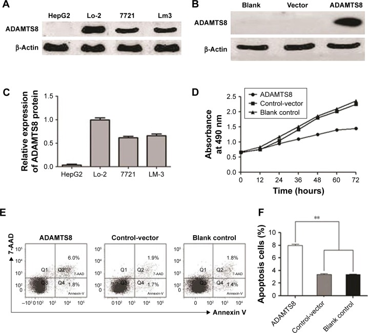
Methods: The tumor tissues and matched adjacent non-tumor tissues were collected from 61 patients with HCC, and ADAMTS8 expression was detected with immunohistochemistry. Flow cytometry and MTT assays were used to assess cell apoptosis and cell viability, respectively, and ERK, p-ERK, Stat3, p-Stat3, Akt, and p-Akt protein expressions were measured by Western blot.

Results: The results showed that ADAMTS8 expression was significantly lower in HCC tissues than that in adjacent non-tumor tissues. Moreover, ADAMTS8 expression was inversely associated with clinical stages and metastasis in patients with HCC. Furthermore, we found that transfection with exogenous ADAMTS8 inhibited proliferation and migration and induced apoptosis in HepG2 cells. In the in vivo study, tumor growth of upregulated HepG2 cells in nude mice was significantly slower. Moreover, decreased ERK activity was detected after transfection with ADAMTS8.

Conclusion: These results indicate that low ADAMTS8 expression is a predictor of a poor prognosis in patients with HCC and that ADAMTS8 plays an important role in regulating HCC growth, invasion, and apoptosis by modulating the ERK signaling pathway. ADAMTS8 maybe a new target in HCC treatment.

Keywords: ADAMT8; apoptosis; hepatocellular carcinoma; invasion and metastasis; signaling pathway.


Conflict of interest statement

Disclosure The authors report no conflicts of interest in this work.

Figures

Figure 1
Figure 1
ADAMTS8 expression in HCC tissues. Notes: ADAMTS8 protein expression was assessed by immunohistochemical analysis. β-Actin was used as a loading control. (A) HCC tissues with ADAMTS8 expression (×200). Black arrow indicates the particular section for the high-resolution image. (B) HCC tissues without ADAMTS8 expression (×200). Abbreviations: ADAMTS, a disintegrin and metallopeptidase with thrombospondin motif; HCC, hepatocellular carcinoma.
Figure 2
Figure 2
The ability of ADAMTS8 to retard HCC cell proliferation in vitro. Notes: (A) ADAMTS8 expression levels in different HCC cell lines. (B) ADAMTS8 protein expression in different HepG2 cell clones was analyzed by Western blotting. Blank group: parental cell lines; Vector group: cells stably transfected with vector alone; ADAMTS8 group: cells stably transfected with vector expressing ADAMTS8. (C) Quantification of results of A. (D) The ability of ADAMTS8 to retard HepG2 cell proliferation was analyzed by MTT assay. (E) The ability of ADAMTS8 to induce apoptosis in HepG2 cells was analyzed by flow cytometry. (F) Quantification of results of E. **P<0.01 compared with the blank control and control-vector groups. Abbreviations: ADAMTS, a disintegrin and metallopeptidase with thrombospondin motif; HCC, hepatocellular carcinoma.
Figure 3
Figure 3
Effects of ADAMTS8 on HCC cell migration and invasion ability. Notes: (A) The effect of ADAMTS8 on HepG2 cell mobility was investigated by wound healing assay. The wounds were photographed, and the migration percentage in a representative experiment was measured using AxioVision software. **P<0.01 compared with the blank control and control-vector groups. (B) Quantification of results of A. (C) Representative transwell and Matrigel assay images and quantification of migrated and invaded cells. *P<0.05, **P<0.01 compared with the blank control and control-vector groups. Abbreviations: ADAMTS, a disintegrin and metallopeptidase with thrombospondin motif; HCC, hepatocellular carcinoma.
Figure 4
Figure 4
Effects of ADAMTS8 on ERK and Stat3/Akt signaling pathway expressions in HepG2 cells. Notes: (A) Western blotting assays of ERK, Stat3, Akt, p-ERK, p-Stat3, and p-Akt protein expressions in HepG2 cells. β-Actin was used as an internal loading control. (B) Western blotting assays of p53 and p21 protein expressions in HepG2 cells. (C) Western blotting assays of p53 and p-ERK protein expressions in vivo. (D) Western blotting assays of ADAMTS8 protein expression in patients with HCC tissues. Line 1, 3, 5: HCC tissues and Line 2, 4, 6: corresponding non-tumor tissues. (E) MTT assay analyzed the ability of ADAMTS8 to retard HepG2 cell proliferation using antitumor drug cisplatin. Abbreviations: ADAMTS, a disintegrin and metallopeptidase with thrombospondin motif; HCC, hepatocellular carcinoma.
Figure 5
Figure 5
Effects of ADAMTS8 on HCC cell growth in vivo. Notes: (A) Xenograft tumor growth curves. (B) The tumor tissues were observed using an animal tissue imaging system. Abbreviations: ADAMTS, a disintegrin and metallopeptidase with thrombospondin motif; HCC, hepatocellular carcinoma.
Figure 6
Figure 6
A schematic diagram of the present finding.

References

    1. Tang BL. ADAMTS: a novel family of extracellular matrix proteases. Int J Biochem Cell Biol. 2001;33(1):33–44. - PubMed
    1. Kumar S, Rao N, Ge R, Rw G. Emerging Roles of ADAMTSs in Angiogenesis and Cancer. Cancers. 2012;4(4):1252–1299. - PMC - PubMed
    1. Mochizuki S, Okada Y. ADAMs in cancer cell proliferation and progression. Cancer Sci. 2007;98(5):621–628. - PMC - PubMed
    1. Rocks N, Paulissen G, El Hour M, et al. Emerging roles of ADAM and ADAMTS metalloproteinases in cancer. Biochimie. 2008;90(2):369–379. - PubMed
    1. Tortorella MD, Burn TC, Pratta MA, et al. Purification and cloning of aggrecanase-1: a member of the ADAMTS family of proteins. Science. 1999;284(5420):1664–1666. - PubMed