Review
. 2018;11(11):26.
doi: 10.1007/s12410-018-9467-z. Epub 2018 Sep 24.

Coronary CTA for Surveillance of Cardiac Allograft Vasculopathy

Affiliations
Review

Coronary CTA for Surveillance of Cardiac Allograft Vasculopathy

Nishant R Shah et al. Curr Cardiovasc Imaging Rep. 2018.

Erratum in

Abstract

Purpose of review: The purpose of this review is to highlight recent hardware and software advances in coronary computed tomography angiography (CTA) that make it a potentially viable alternative to invasive coronary angiography for surveillance of cardiac allograft vasculopathy (CAV) in heart transplant recipients.

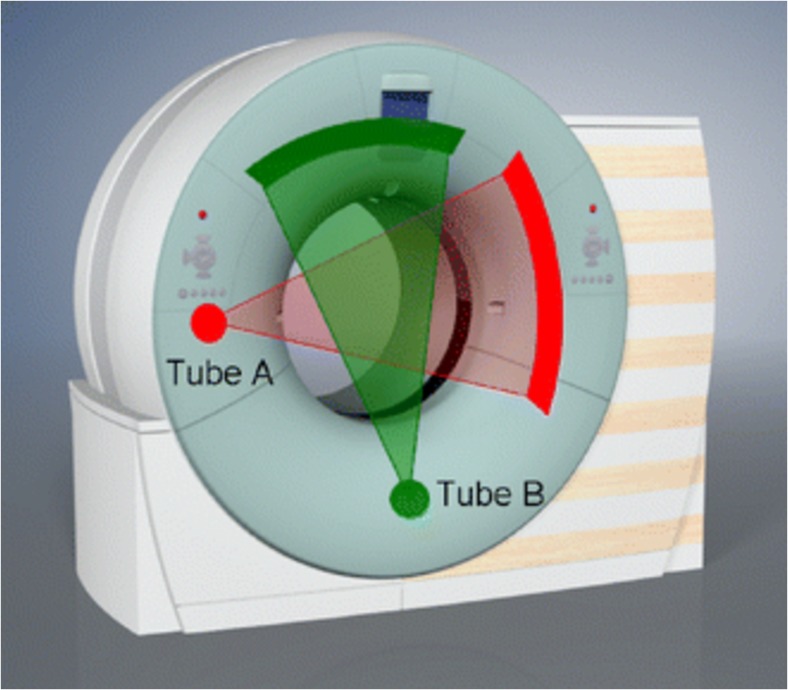
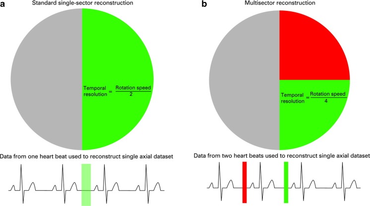
Recent findings: Dual-source CT, multisegment reconstruction, and intracycle motion correction algorithms are all technologies applied during or after image acquisition that can improve image quality and diagnostic accuracy in patients with elevated heart rates, such as heart transplant recipients. CT fractional flow reserve may also add value in this clinical scenario.

Summary: Coronary CTA now has equivalent diagnostic accuracy, offers more nuanced anatomic information, is inherently safer, and could be less costly than invasive coronary angiography. For these reasons, coronary CTA may now be a viable alternative to ICA for CAV surveillance in heart transplant recipients.

Keywords: Cardiac allograft vasculopathy; Coronary CT angiography; Dual-source CT; Intracycle motion correction; Multisegment reconstruction.


Conflict of interest statement

The opinions and assertions herein are those of the authors alone and do not represent the views of the U.S. Army, Office of the Surgeon General, Department of Defense, or the US Government. The authors have reported that they have no relationships relevant to the contents of this paper to disclose.This article does not contain any studies with human or animal subjects performed by any of the authors.

Figures

Fig. 1
Fig. 1
Cardiac allograft vasculopathy (CAV) superimposed on asymmetric native coronary artery disease (CAD; arrowheads). The elastic lamina (open arrow) is intact beneath the CAV lesion but not the CAD lesion. Used with permission from Schoen & Libby, Trends Cardiovasc Med. 1991 Jul-Aug; 1 [5]:216–23
Fig. 2
Fig. 2
Invasive coronary angiogram with intravascular ultrasound images from three representative sites in the left anterior descending coronary artery (LAD) of a 37-year-old man 3 years after heart transplantation. Although the angiogram is without evidence of coronary disease, the ultrasound images demonstrate severe concentric intimal thickening throughout the proximal and mid LAD. Used with permission from St. Goar et al. Circulation. 1992 Mar;85 [3]:979–87
Fig. 3
Fig. 3
Coronary stenosis in the proximal–middle LAD (solid arrows) revealed by both invasive and CT coronary angiography in a heart transplant recipient. Intravascular ultrasounds performed simultaneously confirmed intimal hyperplasia in the proximal LAD (open arrows). Used with permission from Nunoda et al. Circ J. 2010 May;74 [5]:946–53
Fig. 4
Fig. 4
Dual-source computed tomography (DSCT) system with a schematic illustration of the acquisition principle using two tubes and two corresponding detectors offset by 90°. A scanner of this type provides temporal resolution equivalent to a quarter of the gantry rotation time independent of the patient’s heart rate. Used with permission from Flohr et al. Eur Radiol. 2006 Feb;12 [2]:256–68
Fig. 5
Fig. 5
Schematic diagram of single versus multisegment reconstruction showing image acquisition (colored boxes) in mid-late diastole during one vs. two successive cardiac cycles. With multisegment reconstruction (right), image data from multiple cycles are combined to produce the image, thus improving the effective temporal resolution. The circle represents the angular range. Used with permission from Roberts et al. Heart. 2008 Jun;94 [6]:781–792
Fig. 6
Fig. 6
Coronary CTA-derived fractional flow reserve (CT-FFR) in a patient with serial lesions in the left anterior descending coronary artery. Color contours provide data on the distribution of CT-FFR throughout the coronary tree with numerical values obtainable at any location. Used with permission from Taylor et al. J Am Coll Cardiol. 2013 Jun 4;61 [22]:2233–41

References

    1. Lund LH, Khush KK, Cherikh WS, et al. The Registry of the International Society for Heart and Lung Transplantation: thirty-fourth adult heart transplantation report-2017; focus theme: allograft ischemic time. J Heart Lung Transplant. 2017;36:1037–1046. doi: 10.1016/j.healun.2017.07.019. - DOI - PubMed
    1. Salomon RN, Hughes CC, Schoen FJ, Payne DD, Pober JS, Libby P. Human coronary transplantation-associated arteriosclerosis. Evidence for a chronic immune reaction to activated graft endothelial cells. Am J Pathol. 1991;138:791–798. - PMC - PubMed
    1. Mitchell RN, Libby P. Vascular remodeling in transplant vasculopathy. Circ Res. 2007;100:967–978. doi: 10.1161/01.RES.0000261982.76892.09. - DOI - PubMed
    1. Schoen FJ, Libby P. Cardiac transplant graft arteriosclerosis. Trends Cardiovasc Med. 1991;1:216–223. doi: 10.1016/1050-1738(91)90041-C. - DOI - PubMed
    1. Mehra MR, Crespo-Leiro MG, Dipchand A, et al. International Society for Heart and Lung Transplantation working formulation of a standardized nomenclature for cardiac allograft vasculopathy-2010. J Heart Lung Transplant. 2010;29:717–727. doi: 10.1016/j.healun.2010.05.017. - DOI - PubMed

LinkOut - more resources