

Review on Zinc Oxide Nanoparticles: Antibacterial Activity and Toxicity Mechanism
- PMID: 30464967
- PMCID: PMC6223899
- DOI: 10.1007/s40820-015-0040-x
Review on Zinc Oxide Nanoparticles: Antibacterial Activity and Toxicity Mechanism
Abstract
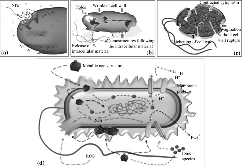
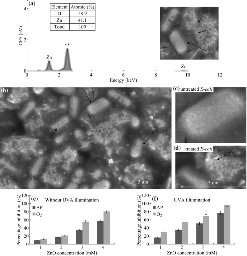
Antibacterial activity of zinc oxide nanoparticles (ZnO-NPs) has received significant interest worldwide particularly by the implementation of nanotechnology to synthesize particles in the nanometer region. Many microorganisms exist in the range from hundreds of nanometers to tens of micrometers. ZnO-NPs exhibit attractive antibacterial properties due to increased specific surface area as the reduced particle size leading to enhanced particle surface reactivity. ZnO is a bio-safe material that possesses photo-oxidizing and photocatalysis impacts on chemical and biological species. This review covered ZnO-NPs antibacterial activity including testing methods, impact of UV illumination, ZnO particle properties (size, concentration, morphology, and defects), particle surface modification, and minimum inhibitory concentration. Particular emphasize was given to bactericidal and bacteriostatic mechanisms with focus on generation of reactive oxygen species (ROS) including hydrogen peroxide (H2O2), OH- (hydroxyl radicals), and O2 -2 (peroxide). ROS has been a major factor for several mechanisms including cell wall damage due to ZnO-localized interaction, enhanced membrane permeability, internalization of NPs due to loss of proton motive force and uptake of toxic dissolved zinc ions. These have led to mitochondria weakness, intracellular outflow, and release in gene expression of oxidative stress which caused eventual cell growth inhibition and cell death. In some cases, enhanced antibacterial activity can be attributed to surface defects on ZnO abrasive surface texture. One functional application of the ZnO antibacterial bioactivity was discussed in food packaging industry where ZnO-NPs are used as an antibacterial agent toward foodborne diseases. Proper incorporation of ZnO-NPs into packaging materials can cause interaction with foodborne pathogens, thereby releasing NPs onto food surface where they come in contact with bad bacteria and cause the bacterial death and/or inhibition.
Keywords: Antibacterial activity; Food antimicrobial; Reactive oxygen species; Toxicity mechanism; Zinc ions release; ZnO-NPs.
Figures
References
-
- S. Sahoo, Socio-ethical issues and nanotechnology development: perspectives from India, in 2010 10th IEEE Conference on Nanotechnology (IEEE-NANO), Seoul, South Korea, USA, 17–20 August 2010 (IEEE, 2010), pp. 1205–1210. doi:10.1109/NANO.2010.5697887
-
- Yadav V. Nanotechnology, big things from a tiny world: a review. AEEE. 2013;3(6):771–778.
-
- B. Ashe, A Detail investigation to observe the effect of zinc oxide and Silver nanoparticles in biological system, M.Sc. (Roll NO-607bm004), National Institute of Technology, 2011
-
- Buzea C, Pacheco II, Robbie K. Nanomaterials and nanoparticles: sources and toxicity. Biointerphases. 2007;2(4):MR17–MR71. - PubMed
Publication types
LinkOut - more resources
Full Text Sources
Other Literature Sources
Research Materials