Review
. 2019 May;137(5):715-730.
doi: 10.1007/s00401-018-1933-9. Epub 2018 Nov 21.

Inflammation in ALS/FTD pathogenesis

Affiliations
Review

Inflammation in ALS/FTD pathogenesis

Madelyn E McCauley et al. Acta Neuropathol. 2019 May.

Abstract

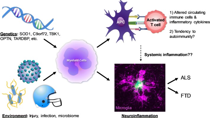
Amyotrophic lateral sclerosis (ALS) and frontotemporal dementia (FTD) are neurodegenerative diseases that overlap in their clinical presentation, pathology and genetics, and likely represent a spectrum of one underlying disease. In ALS/FTD patients, neuroinflammation characterized by innate immune responses of tissue-resident glial cells is uniformly present on end-stage pathology, and human imaging studies and rodent models support that neuroinflammation begins early in disease pathogenesis. Additionally, changes in circulating immune cell populations and cytokines are found in ALS/FTD patients, and there is evidence for an autoinflammatory state. However, despite the prominent role of neuro- and systemic inflammation in ALS/FTD, and experimental evidence in rodents that altering microglial function can mitigate pathology, therapeutic approaches to decrease inflammation have thus far failed to alter disease course in humans. Here, we review the characteristics of inflammation in ALS/FTD in both the nervous and peripheral immune systems. We further discuss evidence for direct influence on immune cell function by mutations in ALS/FTD genes including C9orf72, TBK1 and OPTN, and how this could lead to the altered innate immune system "tone" observed in these patients.

Keywords: Amyotrophic lateral sclerosis; Frontotemporal dementia; Neurodegeneration; Neuroinflammation.


Figures

Fig. 1
Fig. 1
Microglial responses in neuronal injury and ALS/FTD. a, b Immunofluorescence image of Iba1 stain in the region of the motor cortex and corpus callosum (highlighted with dotted white lines) in the normal mouse brain (a), or after experimental traumatic brain injury (b). After tissue injury, Iba1-positive microglia proliferate and become activated with altered morphology, function and transcriptional profile to assist in clearing damage and maintaining tissue homeostasis. Scale bar 200 μm. c Microglial activation in motor cortex from a human subject with sporadic ALS (IBA1—red; DAPI—blue). Microglia appear similar on histology to tissue injury, with proliferation and activation characterized by enlarged ramifications. Scale bar 100 μm. d Iba1 (DAB stain) labeling of ventral horn of a mouse lacking the C9orf72 gene, showing the interaction between the ramifications of a microglial cell and a neighboring neuron, can either be supportive or detrimental in the context of ALS/FTD as discussed in the text. Scale bar 20 μm
Fig. 2
Fig. 2
Schematic of potential interactions between environment, genetics and inflammation in ALS/FTD pathogenesis. Mutations in several ALS/FTD genes can directly disrupt the function of myeloid cells in different compartments and their response to environmental exposures. Altered function of peripheral myeloid-derived antigen-presenting cells (APCs) can promote activation of T cells and B cells, and generate an autoinflammatory state including the production of pro-inflammatory cytokines and autoantibodies, findings that have been observed in patients with ALS. The same genetic mutations can also cause microglial dysfunction, interacting with environmental exposures to drive the pathogenesis of degenerative brain diseases such as ALS/FTD. Finally changes in systemic inflammation can influence the function of microglia and neurons indirectly, and contribute to neurodegeneration. The photomicrograph in the bottom right shows microglia (purple, stained for IBA1) surrounding an amyloid plaque (green, stained for ThioS)

References

    1. Ajami B, Bennett JL, Krieger C, McNagny KM, Rossi FM. Infiltrating monocytes trigger EAE progression, but do not contribute to the resident microglia pool. Nat Neurosci. 2011;14:1142–1149. doi: 10.1038/nn.2887. - DOI - PubMed
    1. Ajami B, Bennett JL, Krieger C, Tetzlaff W, Rossi FM. Local self-renewal can sustain CNS microglia maintenance and function throughout adult life. Nat Neurosci. 2007;10:1538–1543. doi: 10.1038/nn2014. - DOI - PubMed
    1. Akizuki M, Yamashita H, Uemura K, Maruyama H, Kawakami H, Ito H, et al. Optineurin suppression causes neuronal cell death via NF-kappaB pathway. J Neurochem. 2013;126:699–704. doi: 10.1111/jnc.12326. - DOI - PubMed
    1. Alexianu ME, Kozovska M, Appel SH. Immune reactivity in a mouse model of familial ALS correlates with disease progression. Neurology. 2001;57:1282–1289. doi: 10.1212/WNL.57.7.1282. - DOI - PubMed
    1. Alshikho MJ, Zurcher NR, Loggia ML, Cernasov P, Reynolds B, Pijanowski O et al (2018) Integrated MRI and [(11) C]-PBR28 PET imaging in amyotrophic lateral sclerosis. Ann Neurol. https://doi.org/10.1002/ana.25251 - PMC - PubMed

Publication types

MeSH terms