. 2018 Nov 30:9:382-398.
doi: 10.1016/j.isci.2018.11.002. Epub 2018 Nov 4.

PAQR3 Regulates Endoplasmic Reticulum-to-Golgi Trafficking of COPII Vesicle via Interaction with Sec13/Sec31 Coat Proteins

Affiliations

PAQR3 Regulates Endoplasmic Reticulum-to-Golgi Trafficking of COPII Vesicle via Interaction with Sec13/Sec31 Coat Proteins

Qianqian Cao et al. iScience. .

Abstract

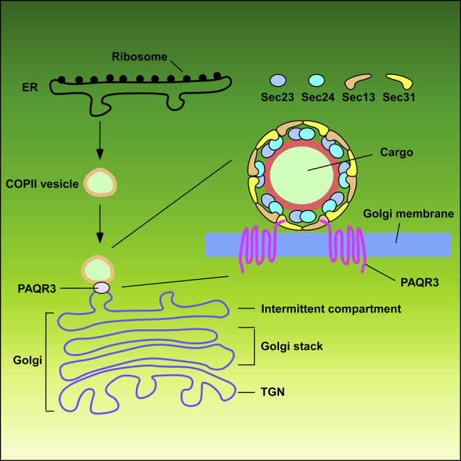
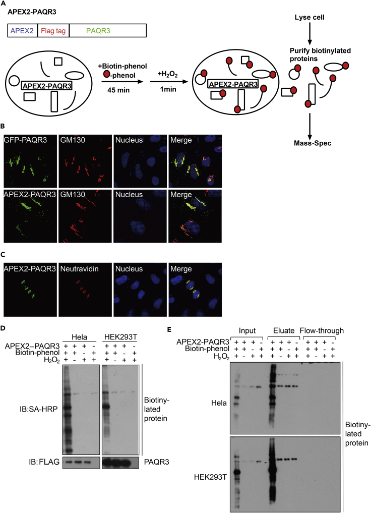
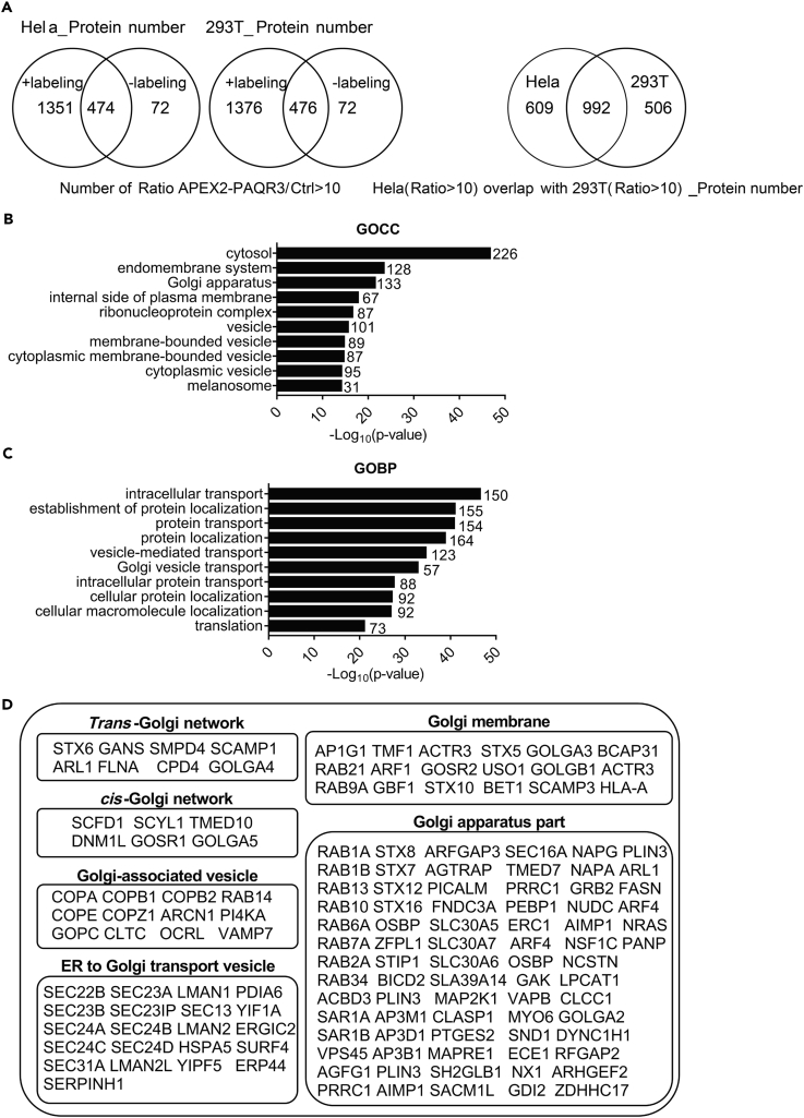
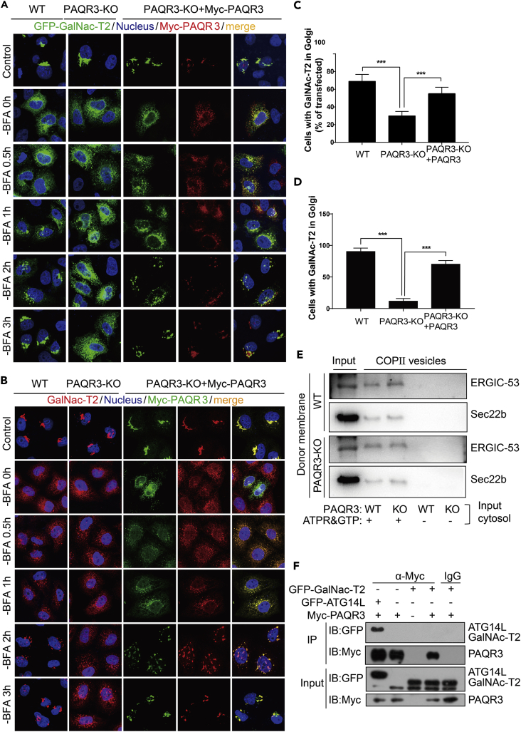
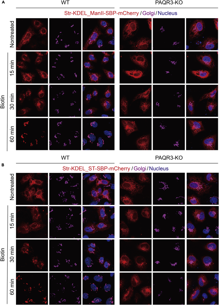
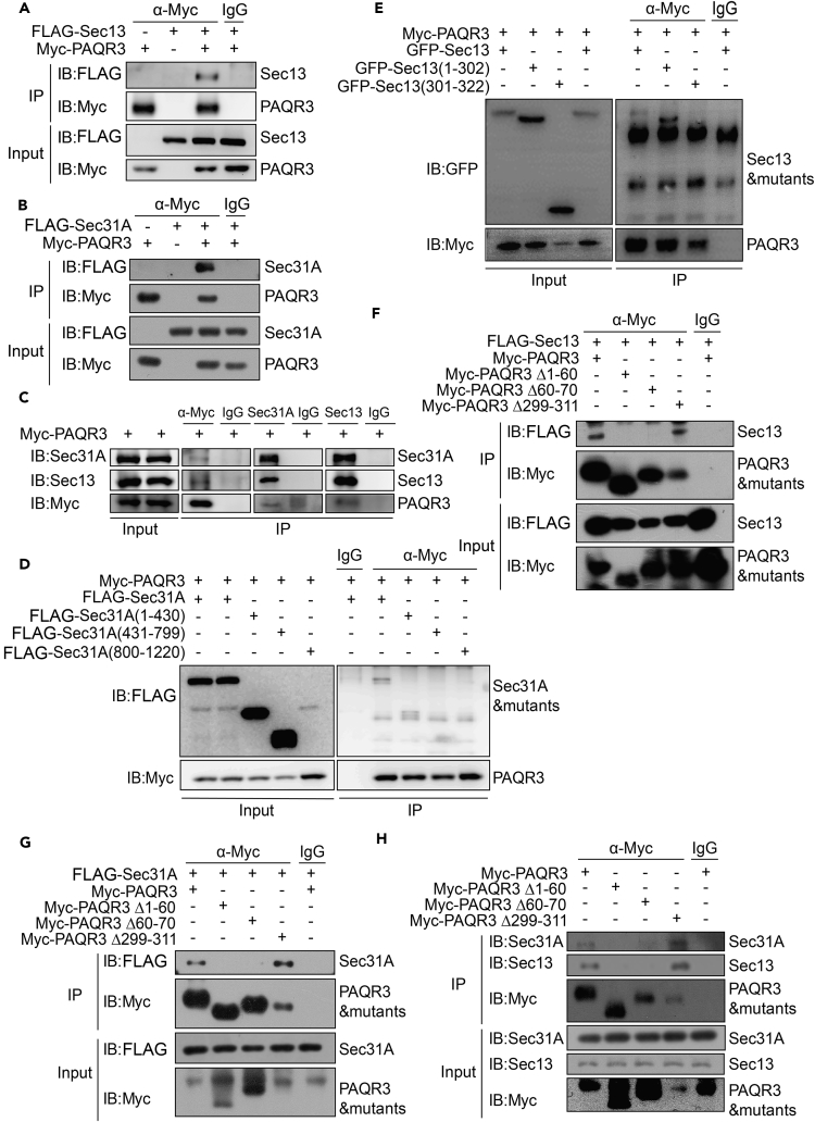
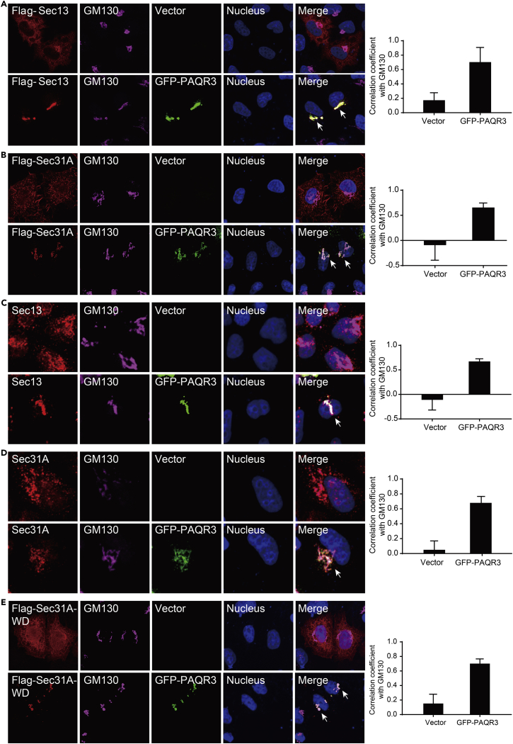
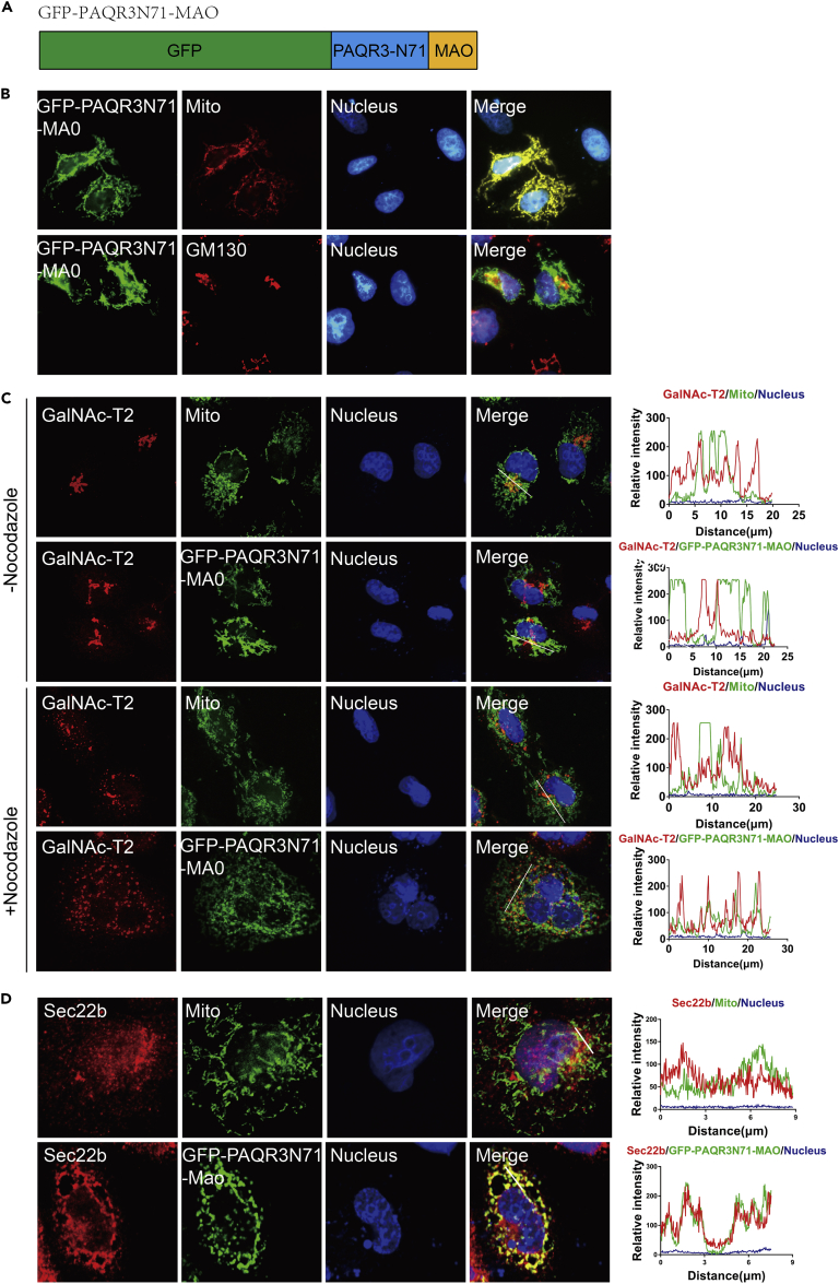
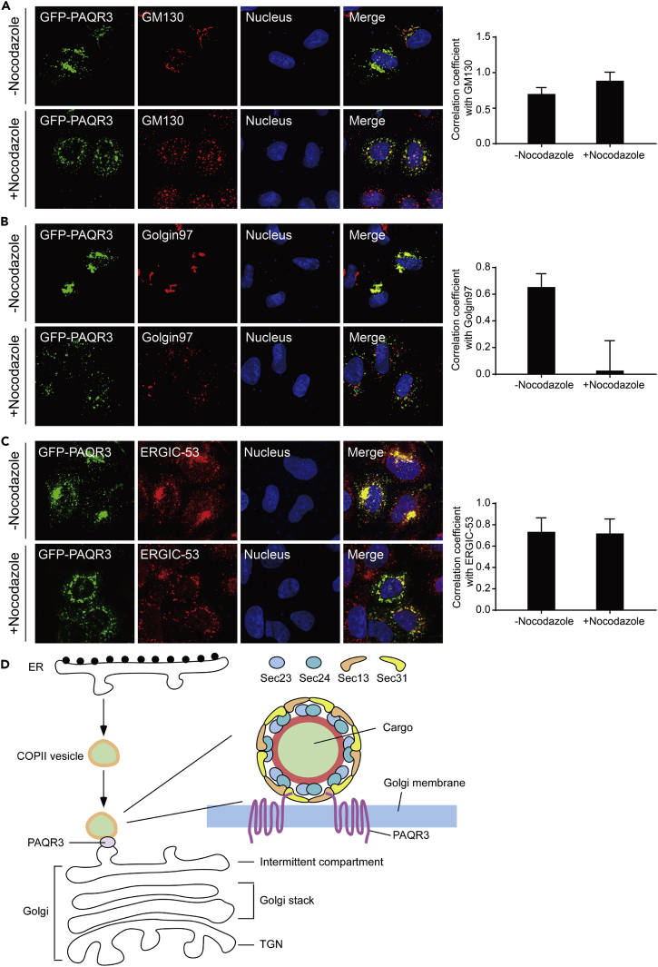
Endoplasmic reticulum (ER)-to-Golgi anterograde transport is driven by COPII vesicles mainly composed of a Sec23/Sec24 inner shell and a Sec13/Sec31 outer cage. How COPII vesicles are tethered to the Golgi is not completely understood. We demonstrated here that PAQR3 can facilitate tethering of COPII vesicles to the Golgi. Proximity labeling using PAQR3 fused with APEX2 identified that many proteins involved in intracellular transport are in close proximity to PAQR3. ER-to-Golgi trafficking of N-acetylgalactosaminyltransferase-2 on removal of brefeldin A is delayed by PAQR3 deletion. RUSH assay also revealed that ER-to-Golgi trafficking is affected by PAQR3. The N-terminal end of PAQR3 can interact with the WD domains of Sec13 and Sec31A. PAQR3 enhances Golgi localization of Sec13 and Sec31A. Furthermore, PAQR3 is localized in the ERGIC and cis-Golgi structures, the acceptor sites for COPII vesicles. Taken together, our study uncovers a role for PAQR3 as a player in regulating ER-to-Golgi transport of COPII vesicles.

Keywords: Cell Biology; Functional Aspects of Cell Biology; Molecular Biology Experimental Approach.


Figures

None
Graphical abstract
Figure 1
Figure 1
Establishment of Proximity Labeling Using APEX2-PAQR3 Fusion Protein (A) Diagram of the APEX2-PAQR3 labeling strategy. APEX2 and a FLAG tag was fused to the N terminus of PAQR3. On sequential treatment with biotin-phenol and H2O2, proteins in close spatial proximity with PAQR3 would be biotinylated (shown as red dots) and then subjected to mass spectrometry analysis. (B) Subcellular distribution of APEX2-fused PAQR3. HeLa cells were transiently transfected with FLAG-tagged APEX2-PAQR3 or GFP-fused PAQR3. The APEX2-PAQR3 was detected by an antibody against FLAG, followed by immunofluorescent staining. The Golgi was stained with an antibody against GM130, and the nucleus was stained with Hoechst 33342. (C) Subcellular distribution of biotinylated proteins. HeLa cells expressing APEX2-PAQR3 were labeled as in (A) and were stained with a NeutrAvidin-Alexa Fluor 647 (AF647) conjugate to visualize biotinylated proteins. An anti-FLAG antibody was used to visualize APEX-PAQR3. (D) Characterization of APEX-PAQR3-mediated biotinylation of endogenous proteins. Hela and HEK293T cells were transfected with APEX2-PAQR3 and treated with or without biotin-phenol/H2O2. The cell lysate was analyzed by immunoblotting with streptavidin-horseradish peroxidase (SA-HRP). The analysis was repeated three times with similar results. (E) Purification of biotinylated proteins. The proteins as in D were purified by streptavidin chromatography, and the samples were analyzed by immunoblotting with SA-HRP.
Figure 2
Figure 2
Identification of Proteins from Proximity Labeling with APEX2-PAQR3 (A) Venn diagram showing identified proteins after streptavidin chromatography from APEX labeling (+labeling) or mock labeling (without H2O2, -labeling) in HeLa cells and HEK293T cells. The right panel shows the proteins found in both cell types. The experiment was repeated three times with similar results. (B) Going analysis of cellular compartment (GOCC) of the 992 candidate proteins found in both HeLa and HEK293K cells. (C) Going analysis of biological process (GOBP) of the 992 candidate proteins found in both cell types. (D) A list of Golgi- and trafficking-associated proteins labeled by APEX2-PAQR3 in both cell types.
Figure 3
Figure 3
Deletion of PAQR3 Delays ER-to-Golgi Trafficking of GalNac-T2 (A and B) ER-to-Golgi transport of exogenous and endogenous GalNAc-T2 in HeLa cells. HeLa cells without or with PAQR3 deletion (WT or PAQR3-KO) were transiently transfected with GFP-GalNAc-T2 and Myc-tagged PAQR3 as indicated. At 24 hr after the transfection, the cells were treated with 5 μg/mL BFA for 30 min. The cells were then analyzed by fluorescence microscopy at different times after BFA removal. Endogenous GalNac-T2 was analyzed by an antibody against GalNAc-T2. The nucleus was stained with Hoechst 33342. All of the images were taken with the same exposure. (C and D) Quantitation of results of A and B. The quantitation of cells with GFP-fused GalNAc-T2 in the Golgi at 3 hr after BFA removal is shown in C. Quantitation of Golgi-localized endogenous GalNAc-T2 at 3 hr after BFA removal is shown in D. At least 500 cells were counted for the quantitation. The data are shown as mean ± SD, *** for p < 0.001. (E) Analysis of COPII vesicle budding in vitro. COPII-budding reactions using cytosolic fractions from WT or PAQR3-KO MEFs and membrane fractions (memb) from WT or PAQR3-KO MEFs. LMAN1/ERGIC-53 and Sec22b are COPII cargos, whose incorporation into budded vesicles needs energy supplied by ATP regeneration system (ATPR) and GTP. The experiment was repeated three times with similar results. (F) Analysis of interaction between PAQR3 and GalNAc-T2. Myc-tagged PAQR3, GFP-fused GalNAc-T2, and GFP-fused ATG14L were transiently expressed in HEK293T cells as indicated. After transfection for 24 hr, the cell lysate was used in immunoprecipitation (IP) and immunoblotting (IB) with the antibodies as indicated. ATG14L was used as a positive control (Xu et al., 2016). The experiment was repeated three times.
Figure 4
Figure 4
PAQR3 Deletion Reduces ER-to-Golgi Trafficking of Golgi-Reporter ST-SBP and ManII-SBP in RUSH Assay Wild-type HeLa cells (WT) or PAQR3-deficient HeLa cells (PAQR3-KO) were transiently transfected with Str-KDEL_ST-SBP-mCherry plasmid or Str-KDEL_ ManII -SBP-mCherry plasmid as indicated. About 36 hr after the transfection, 40 μM of biotin was added for different times. The cells were then analyzed by fluorescence microscopy. The Golgi was stained with antibody against GM130. The nucleus was stained with Hoechst 33342. All of the images were taken with the same exposure. The analysis was repeated three independent times.
Figure 5
Figure 5
PAQR3 Interacts with COPII Components through its N-Terminal Region (A and B) Interaction of PAQR3 with ectopically expressed Sec13 and Sec31A. HEK293T cells were transiently transfected with Myc-tagged PAQR3, FLAG-tagged Sec13, and FLAG-tagged Sec31A as indicated. At 24 hr after the transfection, the cell lysate was used in immunoprecipitation (IP) and immunoblotting (IB) with the antibodies as indicated. (C) Interaction of PAQR3 with endogenous Sec13 and Sec31A. Myc-tagged PAQR3 was expressed in HEK293T cells. After transfection for 24 hr, the cell lysate was used in IP and IB with the antibodies as indicated. (D and E) Identification of the structural domain of Sec13 and Sec31A involved in the interaction with PAQR3. HEK293T cells were transiently transfected with PAQR3 and various mutants of Sec13 and Sec31A as indicated, followed by IP and IB analyses. (F–H) Identification of the domain of PAQR3 involved in the interaction with Sec13 and Sec31A. HEK293T cells were transiently transfected with various PAQR3 mutants and FLAG-tagged Sec13 or FLAG-tagged Sec31A as indicated. The cell lysate was analyzed by IP and IB with the antibodies as indicated. All the western blotting experiments were repeated at least twice with similar results.
Figure 6
Figure 6
PAQR3 Tethers Sec13 and Sec31A to the Golgi Apparatus (A and B) Subcellular localization of exogenous Sec13 and Sec31A in the absence or presence of overexpressed PAQR3. HeLa cells were transiently transfected with FLAG-tagged Sec13, FLAG-tagged Sec31A, and GFP-fused PAQR3 as indicated. The transfected cells were fixed and then stained with an anti-FLAG antibody to detect Sec13/Sec31A. The Golgi was stained with an antibody against GM130, and the nucleus was stained with Hoechst 33342. The arrow indicates apparent co-localization of exogenous Sec13 and Sec31A with PAQR3. (C and D) Subcellular localization of endogenous Sec13 and Sec31A with or without the presence of PAQR3. HeLa cells were transfected with GFP-fused PAQR3 alone. The transfected cells were fixed and then stained with antibodies against Sec13 and Sec31A. The arrow indicates apparent co-localization of endogenous Sec13 and Sec31A with PAQR3. (E) Subcellular localization of the WD domain of Sec31A. HeLa cells were transiently co-transfected with FLAG-tagged WD domain of Sec31A (Sec31A-WD) and GFP-fused PAQR3. The transfected cells were fixed and then stained with an anti-FLAG antibody. The arrow indicates apparent co-localization of the WD domain of Sec31A with PAQR3. Quantitation of the co-localization of the proteins is shown in the right panel (n > 6).
Figure 7
Figure 7
Mitochondria-Localized PAQR3 Alters Localization of GalNAc-T2 to the Mitochondria (A) A diagram depicting the construct of the N-terminal end (N71) of PAQR3 fused with GFP and the C-terminal transmembrane domain of MAO (GFP-PAQR3N71-MAO). (B) Subcellular distribution of GFP-PAQR3N71-MAO. HeLa cells were transiently transfected with GFP-PAQR3N71-MAO, followed by immunofluorescence staining. The mitochondria were stained by MitoTracker Deep Red FM, and the Golgi was stained with an antibody against GM130. The nucleus was stained with Hoechst 33342. (C) GFP-PAQR3N71-MAO changes localization of GalNAc-T2 to the mitochondria. HeLa cells expressing GFP-PAQR3N71-MAO were treated with nocodazole for 6 hr, and endogenous GalNAc-T2 was stained with a specific antibody. The mitochondria were stained by MitoTracker Deep Red FM, and the nucleus was stained with Hoechst 33342. Intensity plots of signal intensity (y axis) against distance in micrometers (x axis) are used to indicate occurrence of overlaps between the red and green channels. (D) GFP-PAQR3N71-MAO changes localization of Sec22b to the mitochondria. HeLa cells were transfected with GFP-PAQR3N71-MAO. Endogenous Sec22b was stained with a specific antibody. The mitochondria were stained by MitoTracker Deep Red FM, and the nuclei were stained with Hoechst 33342. Intensity plots of signal intensity (y axis) against distance in micrometers (x axis) are used to indicate occurrence of overlaps between the red and green channels.
Figure 8
Figure 8
PAQR3 Is Localized in cis-Golgi and ERGIC Structures (A–C) Characterization of PAQR3 localization. HeLa cells were transiently transfected with GFP-fused PAQR3. After transfection for 24 hr, the cells were treated with or without nocodazole for 6 hr to destroy the Golgi, followed by immunofluorescence staining. The cis-Golgi was stained by an antibody against GM130 (A). The trans-Golgi was stained by antibody against Golgin97 (B). The ERGIC structure was stained with an antibody against ERGIC-53(C). The nucleus was stained with Hoechst 33342. Quantitation of the co-localization of the proteins is shown in the right panel (n > 6). (D) A model to depict the function of PAQR3 in regulating COPII trafficking. PAQR3 facilitates tethering of COPII vesicles to ERGIC/cis-Golgi through interaction with the Sec13/Sec31 coat proteins.

Similar articles

Cited by

References

    1. Appenzeller-Herzog C., Hauri H.P. The ER-Golgi intermediate compartment (ERGIC): in search of its identity and function. J. Cell Sci. 2006;119:2173–2183. - PubMed
    1. Aridor M., Bannykh S.I., Rowe T., Balch W.E. Sequential coupling between Copii and Copi vesicle coats in endoplasmic-reticulum to Golgi transport. J. Cell Biol. 1995;131:875–893. - PMC - PubMed
    1. Bannykh S.I., Rowe T., Balch W.E. The organization of endoplasmic reticulum export complexes. J. Cell Biol. 1996;135:19–35. - PMC - PubMed
    1. Bendayan M. Worth its weight in gold. Science. 2001;291:1363–1365. - PubMed
    1. Bersuker K., Peterson C.W.H., To M., Sahl S.J., Savikhin V., Grossman E.A., Nomura D.K., Olzmann J.A. A proximity labeling strategy provides insights into the composition and dynamics of lipid droplet proteomes. Dev. Cell. 2018;44:97–112.e7. - PMC - PubMed

LinkOut - more resources