. 2019 Feb;10(2):482-494.
doi: 10.1016/j.ttbdis.2018.11.006. Epub 2018 Nov 15.

Mutational analysis of gene function in the Anaplasmataceae: Challenges and perspectives

Affiliations

Mutational analysis of gene function in the Anaplasmataceae: Challenges and perspectives

Adela S Oliva Chávez et al. Ticks Tick Borne Dis. 2019 Feb.

Abstract

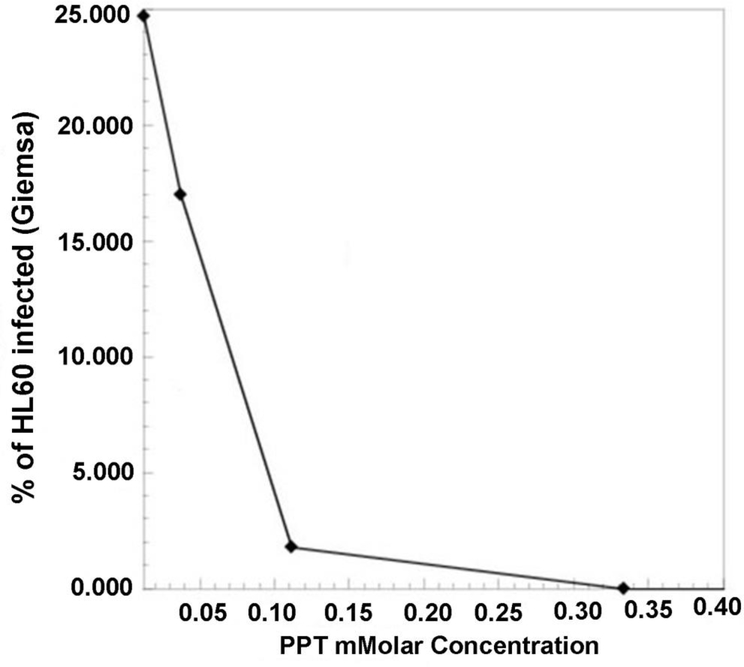
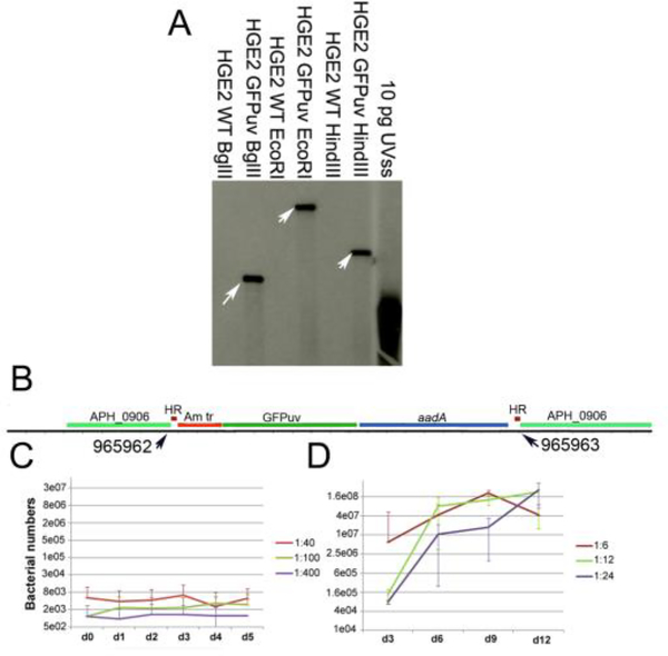
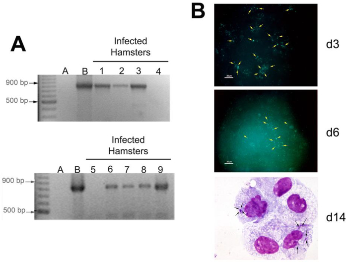
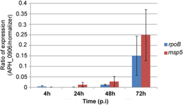
Mutational analysis is an efficient approach to identifying microbial gene function. Until recently, lack of an effective tool for Anaplasmataceae yielding reproducible results has created an obstacle to functional genomics, because surrogate systems, e.g., ectopic gene expression and analysis in E. coli, may not provide accurate answers. We chose to focus on a method for high-throughput generation of mutants via random mutagenesis as opposed to targeted gene inactivation. In our search for a suitable mutagenesis tool, we considered attributes of the Himar1 transposase system, i.e., random insertion into AT dinucleotide sites, which are abundant in Anaplasmataceae, and lack of requirement for specific host factors. We chose the Anaplasma marginale tr promoter, and the clinically irrelevant antibiotic spectinomycin for selection, and in addition successfully implemented non-antibiotic selection using an herbicide resistance gene. These constructs function reasonably well in Anaplasma phagocytophilum harvested from human promyelocyte HL-60 cells or Ixodes scapularis tick cells. We describe protocols developed in our laboratory, and discuss what likely makes them successful. What makes Anaplasmataceae electroporation competent is unknown and manipulating electroporation conditions has not improved mutational efficiency. A concerted effort is needed to resolve remaining problems that are inherent to the obligate intracellular bacteria. Finally, using this approach, we describe the discovery and characterization of a putative secreted effector necessary for Ap survival in HL-60 cells.

Keywords: Anaplasma phagocytophilum; Effector; Selectable markers; Transposon mutagenesis.


Figures

Figure 1.
Figure 1.. Sensitivity of A. phagocytophilum wild-type bacteria grown in HL-60 cells to concentrations of phosphinothricin (PPT).
HL-60 cells infected with wild-type A. phagocytophilum were exposed to increasing concentrations of phosphinothricin (PPT). The number of infected cells was assessed through light microscopy after Giemsa-staining. A concentration of 100 mM PPT was subsequently chosen for selection.
Figure 2.
Figure 2.. Himar single transposition event disrupted the APH_0906 coding gene and affected the growth of A. phagocytophilum in HL-60 cells.
A) Southern blot showing a single insertion site for the himar transposition event. Genomic DNA from ΔAPH_0906 and wild-type bacteria was restriction enzyme digested and hybridized with DIG-probes from the GFPuv gene within the himar transposon. Ten pg of the plasmid containing the original UVSS gene were used as positive control. Arrows point at the single hybridization site within each digestion. B) DNA from ΔAPH_0906 was purified, digested with BglII, and electrophorated into E. coli ElectroMAX DH5α cells. Clone recue was performed and a single colony was sent for sequencing. Sequences were compared against the HZ genome and insertion sites were determined to be at positions 965962 – 965963. C) The growth of the mutant was analyzed q-PCR in HL-60. ΔAPH_0906 mutant was unable to replicate in HL-60 in all three dilutions tested. Red line represents the 1:40, the green line is the 1:100 and the purple line shows the 1:400 dilutions. D) On the other hand, growth within ISE6 cells appeared not to be affected by the mutation. Red line represents the 1:6, the green line is the 1:12 and the purple line shows the 1:24 dilutions.
Figure 3.
Figure 3.. ΔAPH_0906 infects hamsters during in vivo challenge but it is not able to replicate in RF/6A cells in vitro
A) Nine hamsters were challenge with 500 μl of ISE6 cells infected with ΔAPH_0906 bacteria by intraperitoneal (i.p.) injection. Two blood samples were taken 7 days days post-inoculation (p.i.) and the remaining 7 samples were taken at 21 days. DNA was purified from blood samples and nested PCR was used to confirm infection. The two samples taken at day 7 (Lanes 1 and 2) were positive and 5 out of 7 samples were positive at day 21 (Lanes 3–9). DNA from infected cells was used as positive control and the negative control consisted of mice injected with uninfected ISE6 cells. B) RF/6A cells were inoculated with cell free ΔAPH_0906 bacteria. Infections were observed by fluorescence microscopy at day 3 and day 6. Bacteria (pointed by yellow arrows) were observed as green fluorescence associated with cells. This fluorescence appeared to decrease as time passed. Giemsa stains from infected RF/6A at day 14 post-infection (p.i) show very small morulae, suggesting that the bacteria were not able to replicate within the infected cells.
Figure 4.
Figure 4.. APH_0906 mRNA expression within ISE6 and HL-60 cells shows an increase in transcription levels in HL-60 cells.
A) The ratio of expression of the gene encoding the hypothetical protein APH_0906 during infection of HL-60 cells was measured by qRT-PCR at 4, 24, 48, and 72 h p.i. Expression of the gene increased as the infection progressed when normalized against both normalizing genes, rpoB (blue bars) and msp5 (red bars). The ratio of expression was calculated using one delta change of the Ct values (2Δtarget-normalizer). B) The fold change in mRNA transcription of APH_0906 between ISE6 cells and HL-60 cells was measured at same time points described before. The fold change was calculated using the 2−ΔΔct method. As described for the ratio of expression, the transcript levels of APH_0906 increased over time with up-regulation of the gene by 48 h and 72 h of infection when normalized by both genes, rpoB (blue bars) and msp5 (red bars)
Figure 5.
Figure 5.. Subcellular localization of APH_0906 during infection of HL-60 cells.
The subcellular localization of APH_0906 was investigated with Immuno Fluorescence Assays (IFAs) of infected HL-60 cells at 1, 3, and 5 days p.i. The bacteria were labeled with dog serum against A. phagocytophilum detected with FITC anti-dog antibodies (green signal). APH_0906 was labeled using mouse serum raised against recombinant versions of APH_0906 and detected with Cy3-labeled anti-mouse antibodies (red signal). The nucleus of the cells was labeled with DAPI (blue signal). Localization of the protein at day 1 was mostly associated with the bacteria and as infection progressed the protein was translocated into the cytoplasm of the cells by day 3 and 5. A 10 μm size bar is included in the pictures for comparison.
Figure 6.
Figure 6.. Bioinformatic analysis of APH_0906 and its homologs.
A) Phylogenetic relation between homologs of APH_0906 from A. phagocytophilum strains isolated from different hosts analyzed with the UPMAG and B) Neighbor-Joining methods. Strains isolated from human hosts in the US are depicted in dark blue, strains isolated from horses in the US in red, strains isolated from rodents in the US in dark green, strains isolated from ticks in the US in light green, and a strain isolated from a dog in the US is shown in yellow. Strains isolated from dogs in Europe are shown in purple and a strain isolated from a sheep in Norway is shown in light blue. The boostrap values were inferred from 1000 replicates. Three different clusters were identified with dots of different colors at the base of the branches. The red dot identifies a cluster (Cluster 1) formed by APH_0906 homologs from strains isolated from vertebrates in the US. The second cluster (Cluster 2; blue dot) is composed by homologs from strains isolated from vertebrate hosts in Europe, and the last cluster (Cluster 3; green dot) is represented by homologs from strains isolated from ticks in the US. C) MUSCLE alignment of APH_0906 homologs from different clusters showing predicted characteristics within the amino acid sequences. RNA binding sites are represented by pink boxes under the amino acid residues. DNA binding sites are shown in brown and protein binding sites are shown in blue. Predicted Nuclear Localization (NLSs) are shown in orange. Binding sites were predicted with DisoRDPbind and NLSs with NLSmapper and NLStradamus. Image was generated with Geneious R10.

Similar articles

Cited by

References

    1. Absmeier E, Santos KF, Wahl MC, 2016. Functions and regulation of the Brr2 RNA helicase during splicing. Cell Cycle 15, 3362–3377. 10.1080/15384101.2016.1249549 - DOI - PMC - PubMed
    1. Bao Y, Tian M, Li P, Liu J, Ding C, Yu S, 2017. Characterization of Brucella abortus mutant strain Delta22915, a potential vaccine candidate. Vet. Res 48, 17 10.1186/s13567-017-0422-9 - DOI - PMC - PubMed
    1. Barbet AF, Agnes JT, Moreland AL, Lundgren AM, Alleman AR, Noh SM, Brayton KA, Munderloh UG, Palmer GH, 2005. Identification of functional promoters in the msp2 expression loci of Anaplasma marginale and Anaplasma phagocytophilum. Gene 353, 89–97. 10.1016/j.gene.2005.03.036 - DOI - PubMed
    1. Barbet AF, Al-Khedery B, Stuen S, Granquist EG, Felsheim RF, Munderloh UG, 2013. An emerging tick-borne disease of humans is caused by a subset of strains with conserved genome structure. Pathogens 2, 544–555. - PMC - PubMed
    1. Barre N, Happold J, Delathiere J-M, Desoutter D, Salery M, de Vos A, Marchal C, Perrot R, Grailles M, Mortelecque A, 2011. A campaign to eradicate bovine babesiosis from New Caledonia. Ticks Tick. Borne. Dis 2, 55–61. 10.1016/j.ttbdis.2010.11.001 - DOI - PubMed

Publication types

Substances

LinkOut - more resources