. 2018 Nov 22;8(1):17249.
doi: 10.1038/s41598-018-35390-8.

Foxi3 transcription factor activity is mediated by a C-terminal transactivation domain and regulated by the Protein Phosphatase 2A (PP2A) complex

Affiliations

Foxi3 transcription factor activity is mediated by a C-terminal transactivation domain and regulated by the Protein Phosphatase 2A (PP2A) complex

Sunita Singh et al. Sci Rep. .

Abstract

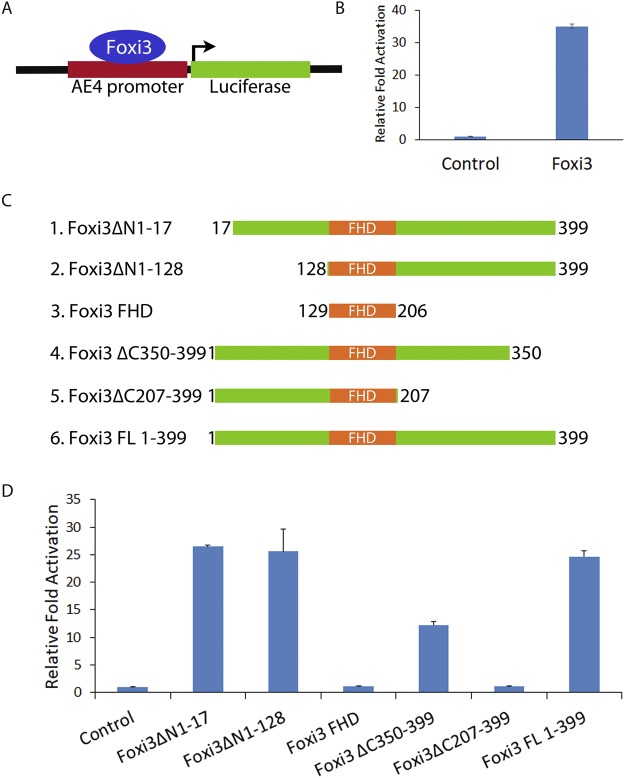
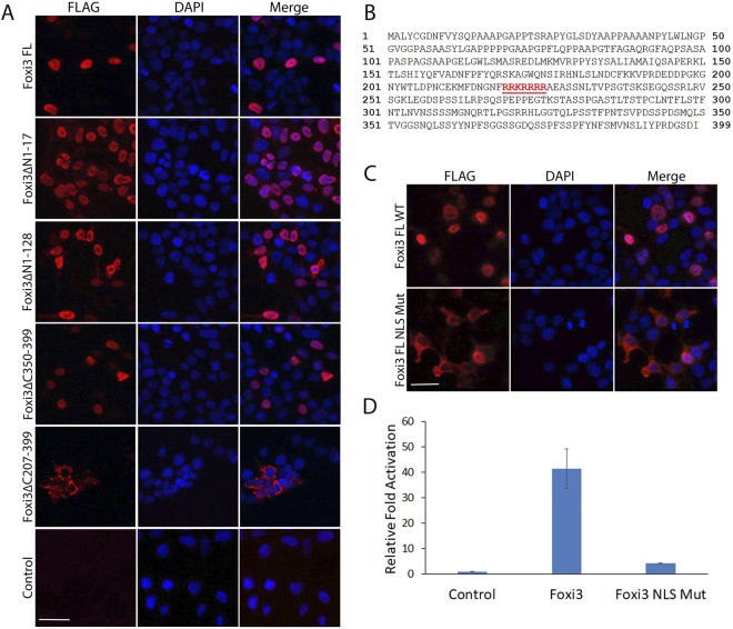
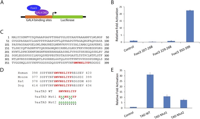
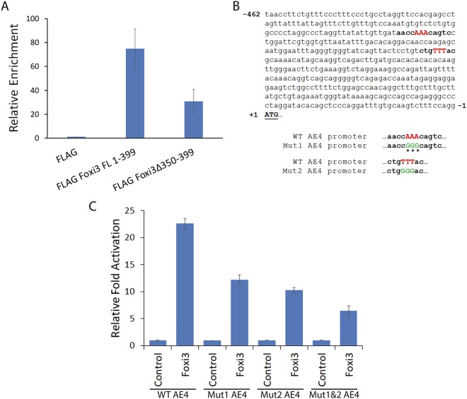
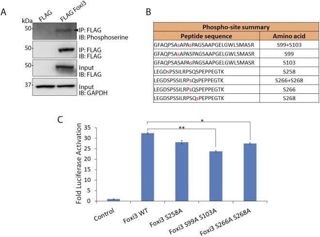
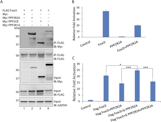
The Forkhead box (FOX) family consists of at least 19 subgroups of transcription factors which are characterized by the presence of an evolutionary conserved 'forkhead' or 'winged-helix' DNA-binding domain. Despite having a conserved core DNA binding domain, FOX proteins display remarkable functional diversity and are involved in many developmental and cell specific processes. In the present study, we focus on a poorly characterized member of the Forkhead family, Foxi3, which plays a critical role in the development of the inner ear and jaw. We show that Foxi3 contains at least two important functional domains, a nuclear localization sequence (NLS) and a C-terminal transactivation domain (TAD), and that it directly binds its targets in a sequence specific manner. We also show that the transcriptional activity of Foxi3 is regulated by phosphorylation, and that the activity of Foxi3 can be attenuated by its physical interaction with the protein phosphatase 2A (PP2A) complex.


Conflict of interest statement

The authors declare no competing interests.

Figures

Figure 1
Figure 1
Identification of functional domains in Foxi3 by deletion analysis. (A) Diagram showing a reporter construct in which the Foxi1-responsive AE4 promoter (22) is cloned upstream of a luciferase reporter gene. (B) Foxi3 activates transcription from the AE4 promoter as shown by a > 30 fold activation of the luciferase reporter. (C) Schematic representation of various N-terminal and C-terminal truncations of Foxi3 which were cloned in 3XFLAG vector. (D) AE4 promoter activity was measured after co-transfection of the AE4 luciferase reporter with various N-terminal and C-terminal truncations of Foxi3. AE4 promoter-linked Luciferase activity is shown as relative fold activation compared with control. Each experiment was performed in triplicate and was repeated at least three times. Error bars represent standard deviations calculated from the biological triplicates. Δ: deletion, FL: full-length, FHD: Forkhead domain.
Figure 2
Figure 2
Characterization of nuclear localization sequence (NLS) in Foxi3. (A) Immunostaining using FLAG antibody in HEK-293T cells transfected with FLAG-tagged N-terminal and C-terminal truncations of Foxi3 described in Fig. 1. DAPI was used as nuclear stain. Deletion of the region from AA207–399 (Foxi3ΔC207–399) prevents nuclear localization. (B) A nuclear localization sequence (219–225aa, shown in red) was predicted for Foxi3 protein using the NucPred tool (C) The predicted NLS was mutated in Foxi3 (Foxi3 FL NLS Mut) and transfected in HEK-293T cells. Wild-type Foxi3 (Foxi3 FL WT) was used as control. Mutation of the predicted NLS abolishes nuclear localization. (D) Mutation of the predicted NLS abolishes Foxi3 activity. AE4 promoter activity was measured after co-transfection of AE4 luciferase reporter with wild-type Foxi3 (Foxi3) or Foxi3 with mutated NLS (Foxi3 NLS Mut). HEK-293T cells were transfected with 250 ng of AE4 luciferase reporter along with 250 ng of either with wild-type Foxi3 (Foxi3) or Foxi3 with mutated NLS (Foxi3 NLS Mut) coding constructs as indicated. AE4 promoter linked Luciferase activity is shown as relative fold activation compared with control. Each experiment was performed in triplicate and was repeated at least three times. Error bars represent standard deviations calculated from the biological triplicates. Δ: deletion, FL: full-length, NLS: nuclear localization sequence. Scale bars: 50 µm.
Figure 3
Figure 3
Characterization and validation of a C-terminal transactivation domain in Foxi3. (A) Foxi3 fragments were cloned as a fusion construct with the DNA binding domain of GAL4 (shown as GAL4 DBD). Foxi3-GAL4 DBD fusion protein binds to the GAL4 binding sites present upstream of a luciferase reporter vector. (B) Luciferase activity was measured after co-transfection of the luciferase reporter with only GAL4 DBD (control) or as a fusion construct of GAL4 DBD with various Foxi3 domains as indicated. Only the C-terminal fragment containing AA350–399 gave significant activation. (C) The Nine Amino Acids Transactivation Domain (9aaTAD) Prediction Tool predicted a 9aa transactivation domain in Foxi3 which is conserved in human, mouse, rat and dog. (D) The conserved 9aaTAD (shown in red) was mutated in the GAL4 DBD-Foxi3 350–399 construct. Two 9aaTAD mutations - 9aaTAD Mut1 and 2 were generated. Mutated aa are shown in green and indicated by an asterisk. (E) Luciferase activity was measured upon co-transfection of the luciferase reporter with only GAL4 DBD (control) or as a fusion construct of GAL4 DBD-Foxi3 350–399 containing wild-type or mutated 9aaTADdomains as indicated. Luciferase activity is shown as relative fold activation compared with control. Each experiment was performed in triplicate and was repeated at least three times. Error bars represent standard deviations calculated from the biological triplicates. DBD: DNA binding domain, TAD: transactivation domain, WT: wild type.
Figure 4
Figure 4
Foxi3 binds directly to the AE4 promoter in a sequence specific manner. (A) Occupancy of the AE4 promoter by Foxi3 was assayed by ChIP analysis. Chromatin was isolated from control (FLAG alone), FLAG Foxi3 FL 1–399 and FLAG Foxi3 Δ350–399 transfected HEK-293T cells and ChIP analysis was performed using Anti-Flag M2 Magnetic Beads. Relative occupancy was calculated by performing quantitative real-time PCR analysis and normalizing the CT values with input and FLAG controls. Each error bar indicates standard deviation calculated from triplicates. (B) The AE4 promoter sequence contains two consensus Forkhead binding motifs ((T/C)AAACA) which are indicated by bold letters. The three ‘AAA’ nucleotides (red) in both consensus sequences were mutated to ‘GGG’ (green) to create two mutant AE4 luciferase reporter constructs labeled as Mut1 and Mut2 AE4 respectively. Mutated nucleotides are shown in green and indicated by the asterisks. We also created a third AE4 luciferase reporter construct where both Mut1 and Mut2 were generated in AE4 promoter and named it as Mut1&2 AE4. (C) Luciferase activity was measured upon co-transfection of the FLAG vector or FLAG Foxi3 with wild-type (WT) AE4, Mut1 AE4 and Mut2 AE4 luciferase reporters. Mutation of one or both Forkhead binding sites significantly decreases reporter activity. HEK-293T cells were transfected with 250 ng of luciferase reporter along with 250 ng of either FLAG (control) or FLAG Foxi3 as indicated. 10 ng of Renilla Luciferase plasmid was used as an internal control in all the transfections. Luciferase activity is shown as relative fold activation compared with control. Each experiment was performed in triplicate and was repeated at least three times. Error bars represent standard deviations calculated from the biological triplicates. FL: full length, Mut: Mutation, WT: wild type.
Figure 5
Figure 5
Foxi3 activity is regulated by phosphorylation. (A) Co-IP showing that Foxi3 is phosphorylated in HEK-293T cells. FLAG-Foxi3 was transfected in HEK-293T cells along with FLAG vector control. Anti-Flag M2 magnetic beads were used to perform immunoprecipitation followed by immunoblotting with anti-phosphoserine. Inputs are shown to validate the expression of FLAG Foxi3, and GAPDH is used to show equal amounts of lysate were used for each immunoprecipitation. Full-length blots are presented in Supplementary Fig. 2. (B) Diagram of the various phosphorylated serine residues identified in Foxi3 by mass spectrometry. (C) Mutation of serine at 99 and 103 amino acid position to alanine results in reduced Foxi3 activity. AE4 promoter activity was measured after co-transfection of AE4 luciferase reporter with wild-type Foxi3 (Foxi3) or Foxi3 with mutated serine residues. HEK-293T cells were transfected with 250 ng of AE4 luciferase reporter along with 250 ng of either with wild-type Foxi3 (Foxi3) or Foxi3 serine mutant coding constructs as indicated. AE4 promoter linked luciferase activity is shown as relative fold activation compared with control. Each experiment was performed in triplicate and was repeated at least three times. Error bars represent standard deviations calculated from the biological triplicates. Statistical significance was determined by Student’s t test. A value of P < 0.05 was considered as statistically significant (*P < 0.05; **P < 0.005). IB: Immunoblotting; IP: Immunoprecipitation; kDa: kiloDalton.
Figure 6
Figure 6
Foxi3 is negatively regulated by its interaction with the PP2A complex. (A) Co-IP showing the interaction between Foxi3 and various PP2A subunits. Myc-tagged PP2A subunits, PPP2R2A, PPP2CB and PPP2R1A were transfected along with FLAG Foxi3. Anti-Flag M2 magnetic beads were used to perform immunoprecipitations and MYC was used to detect the PP2A subunits. Inputs are shown to validate the expression of indicated constructs and GAPDH is used to show equal amounts of lysate were used for each immunoprecipitation. Full-length blots are presented in Supplementary Fig. 2. (B) Reporter assay showing that co-expression of PPP2R2A reduces the Foxi3-mediated activation of the AE4 luciferase reporter. PPP2R2A itself does not have any effect on the reporter activity. (C) Expression of exogenous PPP2R2A attenuates Foxi3 reporter activation. In contrast, Foxi3-mediated reporter activation is increased by knockdown of endogenous PPP2R2A in HEK-293T cells. The effect of siRNA knockdown of PPP2R2A on Foxi3 is reversed by overexpression of a PPP2R2A cDNA. Each experiment was performed in triplicate and was repeated at least three times. P-values are calculated Results are expressed as the mean ± SD calculated from triplicates. Statistical significance was determined by Student’s t test. A value of P < 0.05 was considered as statistically significant (*P < 0.05; ***P < 0.0005). IB: Immunoblotting; IP: Immunoprecipitation; kDa: kiloDalton.

Similar articles

Cited by

References

    1. Myatt SS, Lam EW. The emerging roles of forkhead box (Fox) proteins in cancer. Nat Rev Cancer. 2007;7:847–859. doi: 10.1038/nrc2223. - DOI - PubMed
    1. Lam EW, Brosens JJ, Gomes AR, Koo CY. Forkhead box proteins: tuning forks for transcriptional harmony. Nat Rev Cancer. 2013;13:482–495. doi: 10.1038/nrc3539. - DOI - PubMed
    1. Golson ML, Kaestner KH. Fox transcription factors: from development to disease. Development. 2016;143:4558–4570. doi: 10.1242/dev.112672. - DOI - PMC - PubMed
    1. Lai E, et al. HNF-3A, a hepatocyte-enriched transcription factor of novel structure is regulated transcriptionally. Genes Dev. 1990;4:1427–1436. doi: 10.1101/gad.4.8.1427. - DOI - PubMed
    1. Weigel D, Jackle H. The fork head domain: a novel DNA binding motif of eukaryotic transcription factors? Cell. 1990;63:455–456. doi: 10.1016/0092-8674(90)90439-L. - DOI - PubMed

Publication types