

Effects of Low Intensity Focused Ultrasound on Liposomes Containing Channel proteins
- PMID: 30467339
- PMCID: PMC6250712
- DOI: 10.1038/s41598-018-35486-1
Effects of Low Intensity Focused Ultrasound on Liposomes Containing Channel proteins
Abstract
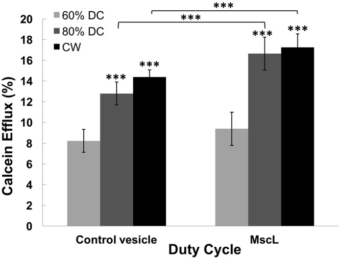
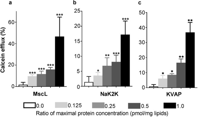
The ability to reversibly and non-invasively modulate region-specific brain activity in vivo suggests Low Intensity Focused Ultrasound (LIFU) as potential therapeutics for neurological dysfunctions such as epilepsy and Parkinson's disease. While in vivo studies provide evidence of the bioeffects of LIFU on neuronal activity, they merely hint at potential mechanisms but do not fully explain how this technology achieves these effects. One potential hypothesis is that LIFU produces local membrane depolarization by mechanically perturbing the neuronal cell membrane, or activating channels or other proteins embedded in the membrane. Proteins that sense mechanical perturbations of the membrane, such as those gated by membrane tension, are prime candidates for activating in response to LIFU and thus leading to the neurological responses that have been measured. Here we use the bacterial mechanosensitive channel MscL, which has been purified and reconstituted in liposomes, to determine how LIFU may affect the activation of this membrane-tension gated channel. Two bacterial voltage-gated channels, KvAP and NaK2K F92A channels were also studied. Surprisingly, the results suggest that ultrasound modulation and membrane perturbation does not induce channel gating, but rather induces pore formation at the membrane protein-lipid interface. However, in vesicles with high MscL mechanosensitive channel concentrations, apparent decreases in pore formation are observed, suggesting that this membrane-tension-sensitive protein may serve to increase the elasticity of the membrane, presumably because of expansion of the channel in the plane of the membrane independent of channel gating.
Conflict of interest statement
The authors declare no competing interests.
Figures

References
-
- Vonck K, et al. Neurostimulation for refractory epilepsy. Acta neurologica belgica. 2003;103:212–217. - PubMed
-
- Engel J, et al. Practice parameter: Temporal lobe and localized neocortical resections for epilepsy Report of the Quality Standards Subcommittee of the American Academy of Neurology, in Association with the American Epilepsy Society and the American Association of Neurological Surgeons. Neurology. 2003;60:538–547. doi: 10.1212/01.WNL.0000055086.35806.2D. - DOI - PubMed
-
- Fitzgerald, P. B. & Daskalakis, Z. J. The effects of repetitive transcranial magnetic stimulation in the treatment of depression (2011). - PubMed
