Review
. 2019 Jan;20(1):19-33.
doi: 10.1038/s41583-018-0093-1.

Necroptosis and RIPK1-mediated neuroinflammation in CNS diseases

Affiliations
Review

Necroptosis and RIPK1-mediated neuroinflammation in CNS diseases

Junying Yuan et al. Nat Rev Neurosci. 2019 Jan.

Abstract

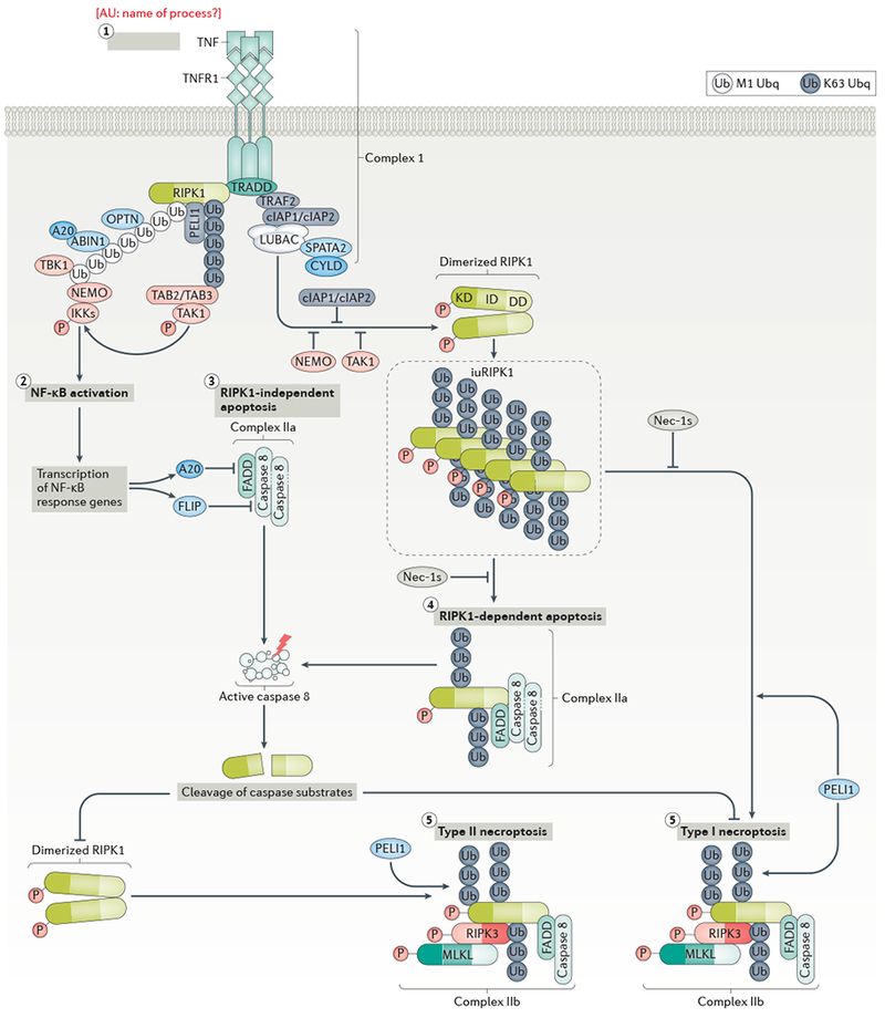
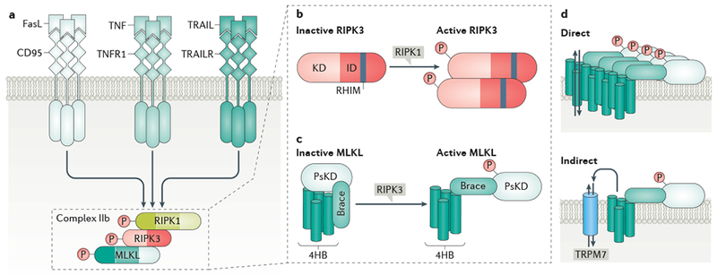
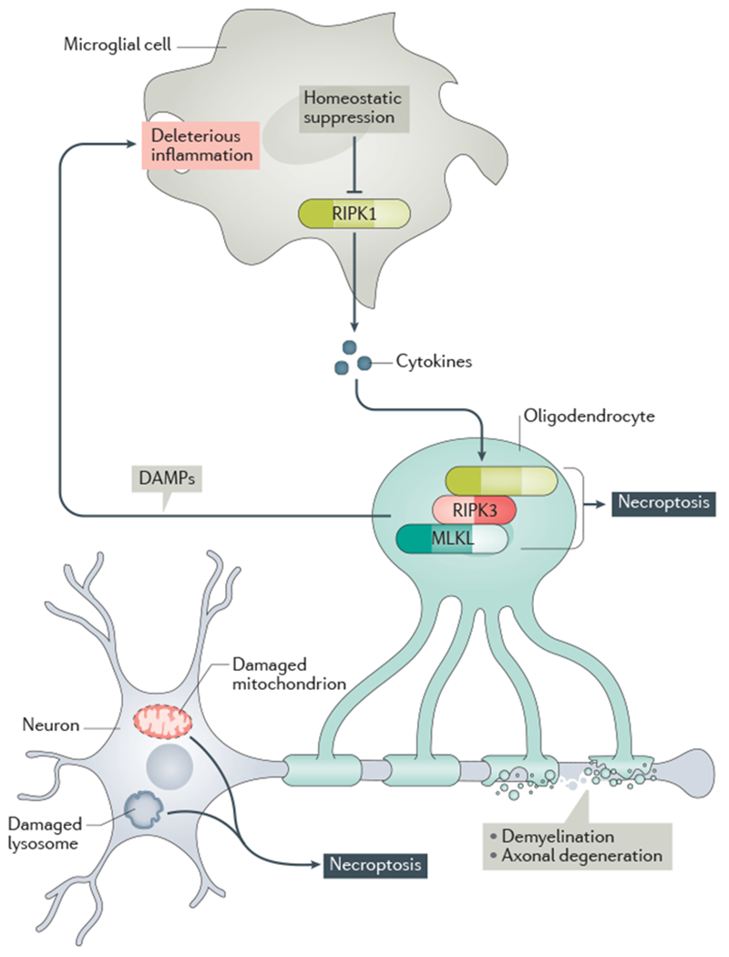
Apoptosis is crucial for the normal development of the nervous system, whereas neurons in the adult CNS are relatively resistant to this form of cell death. However, under pathological conditions, upregulation of death receptor family ligands, such as tumour necrosis factor (TNF), can sensitize cells in the CNS to apoptosis and a form of regulated necrotic cell death known as necroptosis that is mediated by receptor-interacting protein kinase 1 (RIPK1), RIPK3 and mixed lineage kinase domain-like protein (MLKL). Necroptosis promotes further cell death and neuroinflammation in the pathogenesis of several neurodegenerative diseases, including multiple sclerosis, amyotrophic lateral sclerosis, Parkinson disease and Alzheimer disease. In this Review, we outline the evidence implicating necroptosis in these neurological diseases and suggest that targeting RIPK1 might help to inhibit multiple cell death pathways and ameliorate neuroinflammation.


Conflict of interest statement

Competing interests

J.Y. is a consultant of Denali Therapeutics. P.A. declares no competing interests. D.O. is an employee of Sanofi.

Figures

Fig. 1 |
Fig. 1 |. TNF signalling-mediated apoptosis and necroptosis.
a | The binding of tumour necrosis factor (TNF) to TNF receptor 1 (TNFR1) leads to receptor trimerization and activation. TNFR1-associated death domain (DD) protein (TRADD) and receptor-interacting protein kinase 1 (RIPK1) are recruited to the intracellular DD of activated TNFR1 via their own DDs to initate the formation of complex I. In complex I, TRADD recruits the E3 ubiquitin ligases cellular inhibitor of apoptosis 1 (cIAP1) and cIAP2, which in turn add K63 ubiquitin chains to complex I members, including to the K377 residue of RIPK1. The linear ubiquitylation assembly complex (LUBAC) is recruited by binding with K63 ubiquitin chains in complex I and performs linear (M1) ubiquitylation of RIPK1. Deubiquitinating enzyme CYLD and its adaptor protein SPATA2 are recruited together with the LUBAC to complex I. Transforming-growth-factor-β-activated kinase 1 (TAK1) is recruited to K63 ubiquitin chains on RIPK1 via TAK1-binding protein 2 (TAB2) or TAB3. The nuclear factor-κB (NF-κB) essential modulator (NEMO)–IκB kinase (IKK) complex and the kinase TBK1 are recruited to complex I by binding with M1 ubiquitin chains on RIPK1. M1-ubiquitin binding proteins ABIN-1 and OPTN are also recruited to complex I. ABIN-1 in turn recruits ubiquitin editing enzyme A20 to complex I. b | Under normal conditions, activated IKKs and TAK1 lead to NF-κB activation and transcription of pro-inflammatory and pro-survival genes, such as those encoding FLICE-inhibitory protein (FLIP) and A20, which suppress the activation of caspase-8 and RIPK1, respectively. c | When NF-κB activation is inhibited, a cytosolic complex named complex IIa can form to mediate the activation of caspase-8 and subsequent cleavage of RIPK1 and RIPK1-independent apoptosis. d | The cleavage of RIPK1 at the C-terminus of its kinase domain (after residue D324) by caspase 8 provides an important mechanism by which apoptosis, both RIPK1-dependent and RIPK1-independent, may pre-empt the activation of RIPK1 and necroptosis. When caspase-8 activation is blocked, dimerization of RIPK1 via its C-terminal DD leads to RIPK1 activation and formation of complex IIb, which includes FADD, caspase-8, RIPK1, RIPK3 and MLKL.. RIPK1 activity is required for the formation of complex IIb. PELI1 mediates K63 ubiquitination of RIPK1 in complex IIb which promotes the activation of RIPK3 and MLKL to mediate type II necroptosis. e | Alternatively, when suppression of RIPK1 activity in complex I fails under pathological conditions, RIPK1 may mediate RIPK1-dependent apoptosis or type I necroptosis. Specifically, under cIAP1/2, NEMO-, IKK- or TAK1-deficient conditions, an activated, detergent-insoluble ubiquitinated RIPK1 species (iuRIPK1) may form following complex I to accelerate RIPK1 dimerization and subsequent formation of complex II and RIPK1-dependent apoptosis or type I necroptosis. f | When caspase-8 activation is blocked, the formation of complex IIb, which includes RIPK1, RIPK3, and MLKL, can lead to type I necroptosis. High levels of activated RIPK1 in complex I and the formation of iuRIPK1 distinguish type I necroptosis from type II necroptosis.Inhibition of RIPK1 kinase by Nec-1s blocks RIPK1 activation to prevent RDA and necroptosis.
Fig. 2 |
Fig. 2 |. Post-translational modifications regulate the activation of RIPK1.
The domain structure of receptor-interacting protein 1 (RIPK1) consists of an N-terminal kinase domain, an intermediate domain and a C-terminal death domain (DD). The N-terminal kinase domain is crucial for RIPK1-dependent apoptosis and necroptosis. The cleavage of RIPK1 at D324 by caspase-8 blocks the activation of RIPK1 by separating the kinase domain from the C-terminal DD, which is critical for mediating RIPK1 activation by dimerization. Autophosphorylation of Ser14/15 and Ser166 of RIPK1 are biomarkers of its kinase activation. K115 in activated RIPK1 is ubiquitylated by the E3 ubiquitin ligase pellino 1 (PELI1). The kinase TBK1 phosphorylates T189 RIPK1 to suppress its activation. The intermediate domain of RIPK1 is involved in regulating nuclear factor-κB (NF-κB) signalling. RIPK1 can be ubiquitylated on multiple residues within this domain; one of these sites, Lys377, is a crucial residue that can be ubiquitylated by E3 ubiquitin ligases such as cellular inhibitor of apoptosis 1 (cIAP1) or cIAP2 and parkin. The Lys377 residue also serves as a binding hub for downstream signalling, including the NF-κB signalling pathway, by recruiting K63 ubiquitin binding proteins TAK1-binding protein 2 (TAB2) or TAB3, which in turn promote the activation of transforming-growth-factor-β-activated kinase 1 (TAK1). Activated TAK1 mediates multiple inhibitory phosphorylations of RIPK1 directly and indirectly by promoting the activation of IκB kinases (IKKs) and MAPK-activated protein kinase 2 (MK2). TBK1 mediates inhibitory phosphorylation of RIPK1 on T189 to block its substrate recognition in autophosphorylation-mediated activation. The receptor-interacting protein homotypic interaction motif (RHIM) within the intermediate domain of RIPK1 mediates homotypic binding with the RHIM of RIPK3 to promote downstream signalling in necroptosis and inflammation. The C-terminal DD of RIPK1 mediates heterodimerization with the death domains of TNFR1, TRADD and FADD, as well as homodimerization with itself to mediate the activation of N-terminal kinase domain. M1 ubiquitylation of RIPK1 by the linear ubiquitylation assembly complex (LUBAC) suppresses RIPK1 activation by enabling the recruitment of multiple UBAN-containing ubiquitin-binding proteins, including NEMO, ABIN1 and OPTN, which promote the activation of IKKs (NEMO), recruit A20 (ABIN1) or regulate K48 ubiquitylation of RIPK1 (OPTN).
Fig. 3 |
Fig. 3 |. Execution of necroptosis.
a | Activation of death receptors such as FAS, tumour necrosis factor (TNF) receptor 1 (TNFR1) or TNF-related apoptosis-inducing ligand receptors (TRAILRs) by their respective ligands can lead to the RIPK1 dimerization and activation, which in turn promotes the formation of a RIPK1–RIPK3–MLKL complex, referred to as complex IIb, and activation of RIPK3 and MLKL. b | RIPK3 consists of an N-terminal kinase domain and a C-terminal intermediate domain containing a RHIM motif that mediates binding to RIPK1. Dimerization or oligomerization of RIPK3, through RIPK1 kinase-dependent complex IIb formation, allows transphosphorylation of RIPK3 at T231/S232 in mouse RIPK3 and S227 in human RIPK3, and RIPK3 activation. c | MLKL contains a N-terminal 4-helix bundle (4HB), a brace region and a C-terminal pseudo-kinase domain (PsKD). Phosphorylation of the PsKD of MLKL by RIPK3, at S345 in mouse MLKL or T357/S358 in human MLKL, results in a conformational change of MLKL to reveal phosphatidylinositol phosphate (PIP) binding sites. d | Phosphorylated MLKL binds directly to PIPs and cardiolipin in the plasma membrane or cellular organelle membranes, leading to membrane disruption and cell lysis. The precise mechanism is unclear, but this may occur directly through MLKL oligomerization and pore formation, or indirectly, through activation of transporters or ion channels, such as transient receptor potential cation channel M7 (TRPM7).
Fig. 4 |
Fig. 4 |. Bimodal deleterious RIPK1 activation in neurological disease.
Microglial RIPK1 regulates a degenerative neuroinflammatory milieu in the CNS that can lead to necroptosis of oligodendrocytes and axonal degeneration. In neurons, damaged mitochondria and lysosomes can promote necroptosis. In turn, necroptosis can promote inflammation by driving the cell-autonomous expression of proinflammatory cytokines as well as by releasing of the cellular content from necrotic cells (including damage-associated molecular patterns (DAMPs)) into the CNS. This deleterious axis creates a progressive inflammatory and degenerative environment in the brain to promote the progression of neurodegenerative disease.

References

    1. Yuan J & Yankner BA Apoptosis in the nervous system. Nature 407, 802–809, doi:10.1038/35037739 (2000). - DOI - PubMed
    1. Degterev A et al. Chemical inhibitor of nonapoptotic cell death with therapeutic potential for ischemic brain injury. Nat Chem Biol 1, 112–119 (2005). - PubMed
    2. This paper provided the first definition of necroptosis and insights into the functional role of necroptosis in acute neurological injuries.

    1. Degterev A et al. Identification of RIP1 kinase as a specific cellular target of necrostatins. Nat Chem Biol 4, 313–321 (2008). - PMC - PubMed
    2. This paper demonstrated RIPK1 kinase as the target of necrostatin-1 and the role of RIPK1 as a key mediator of necroptosis.

    1. Ofengeim D & Yuan J Regulation of RIP1 kinase signalling at the crossroads of inflammation and cell death. Nat Rev Mol Cell Biol 14, 727–736, doi:10.1038/nrm3683 (2013). - DOI - PubMed
    1. Weinlich R, Oberst A, Beere HM & Green DR Necroptosis in development, inflammation and disease. Nat Rev Mol Cell Biol 18, 127–136, doi:10.1038/nrm.2016.149 (2017). - DOI - PubMed

Publication types

Substances