. 2019 Feb;21(1):111-123.
doi: 10.1007/s10126-018-9865-x. Epub 2018 Nov 22.

Genetic Mutations in jamb, jamc, and myomaker Revealed Different Roles on Myoblast Fusion and Muscle Growth

Affiliations

Genetic Mutations in jamb, jamc, and myomaker Revealed Different Roles on Myoblast Fusion and Muscle Growth

Yufeng Si et al. Mar Biotechnol (NY). 2019 Feb.

Abstract

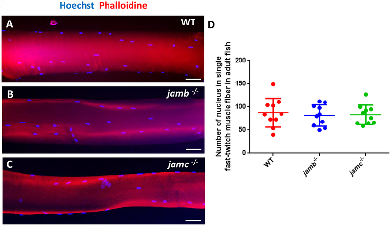
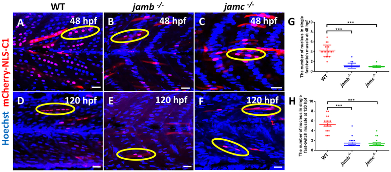
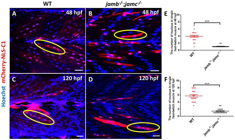
Myoblast fusion is a vital step for skeletal muscle development, growth, and regeneration. Loss of Jamb, Jamc, or Myomaker (Mymk) function impaired myoblast fusion in zebrafish embryos. In addition, mymk mutation hampered fish muscle growth. However, the effect of Jamb and Jamc deficiency on fish muscle growth is not clear. Moreover, whether jamb;jamc and jamb;mymk double mutations have stronger effects on myoblast fusion and muscle growth remains to be investigated. Here, we characterized the muscle development and growth in jamb, jamc, and mymk single and double mutants in zebrafish. We found that although myoblast fusion was compromised in jamb and jamc single or jamb;jamc double mutants, these mutant fish showed no defect in muscle cell fusion during muscle growth. The mutant fish were able to grow into adults that were indistinguishable from the wild-type sibling. In contrast, the jamb;mymk double mutants exhibited a stronger muscle phenotype compared to the jamb and jamc single and double mutants. The jamb;mymk double mutant showed reduced growth and partial lethality, similar to a mymk single mutant. Single fiber analysis of adult skeletal myofibers revealed that jamb, jamc, or jamb;jamc mutants contained mainly multinucleated myofibers, whereas jamb;mymk double mutants contained mostly mononucleated fibers. Significant intramuscular adipocyte infiltration was found in skeletal muscles of the jamb;mymk mutant. Collectively, these studies demonstrate that although Jamb, Jamc, and Mymk are all involved in myoblast fusion during early myogenesis, they have distinct roles in myoblast fusion during muscle growth. While Mymk is essential for myoblast fusion during both muscle development and growth, Jamb and Jamc are dispensable for myoblast fusion during muscle growth.

Keywords: Jamb; Jamc; Muscle fusion; Myomaker; Zebrafish.


Conflict of interest statement

Conflict of Interest Statement

All authors declared no conflict of interest.

Figures

Fig. 1.
Fig. 1.. Loss of jamb or jamc had no effect on fish muscle development and growth
A-C: Morphological comparison of WT control (A), jamb (B) or jamc (C) mutant fish at 7 months old. Scale bar (500 mm). D-I: HE staining showing cross sections of trunk skeletal muscles in WT control (D, G), jamb (E, H) or jamc (F, I) mutant fish of 7 months old. Scale bar (250 μm).
Fig. 2.
Fig. 2.. Single fiber analysis of myonuclei in skeletal muscles from adult WT, jamb and jamc mutants
Single fibers were isolated from skeletal muscles of WT adult fish (A), jamb (B) or jamc (C) mutants at 7 months old. The isolated single fibers were stained with Hoechst 32258 and phalloidin-TRITC, and photographed under a confocal microcopy. Statistical analysis was performed to compare the numbers of nucleus in multiple myofibers from the three different groups. No significant difference was detected in myofibers from these three groups of fish. Scale bar (50 μm).
Fig. 3.
Fig. 3.. Characterization of myoblast fusion in jamb or jamc mutant embryos
Myoblast fusion was analyzed in WT, jamb or jamc mutants by nuclear labelling with Hoechst 32258 (nuclei, blue) and nuclear mCherry (mCherry-NLS-C1; red) at 48 (A-C) and 120 (D-F) hpf. A: WT control contained mostly multinucleated fast myofibers at 48 hpf. B, C: Myofibers in jamb (B) or jamc (C) mutant were mostly mononucleated fibers with a central nuclear localization within the myotome. However, a small number of binucleated myofibers were observed in the jamb and jamc mutants. D: WT control contained mostly multinucleated fast myofibers at 120 hpf. E, F: A broader distribution of myonuclei was detected in the myotome of jamb (E) and jamc (F) mutants at 120 hpf. G, H: Statistical analysis was performed to compare the numbers of nucleus in myofibers from multiple embryos for each genotype (n=20). Significant difference (p < 0.05) was observed between WT and jamb or jamc mutant, whereas no significant difference was detected between jamb and jamc mutants. Scale bar (25μm).
Fig. 4.
Fig. 4.. The effect of jamb and jamc double mutation on myoblast fusion in zebrafish embryos
The mCherry-NLS-C1 plasmid was injected into WT control and jamb;jamc double mutant embryos at 1–2 cell stages. The injected embryos were fixed at 48 and 120 hpf. Myonuclei were labelled with Hoechst 32258 (blue) and mCherry-NLS-C1 (red). A, C: Myofibers in WT embryos contained multiple nuclei per fiber at 48 (A) and 120 (C) hpf. B, D: Myofibers in jamb;jamc double mutants were mostly mononucleated fibers at 48 (B) and 120 (D) hpf. However, a few myofibers containing 2–3 myonuclei were detected in the jamb;jamc double mutants at 48 and 120 hpf. E, F: *** showed the significant statistical difference between WT and jamb;jamc double mutant when comparing the number of nucleus in single fast-twitch muscle fiber at 48 (E) and 120 (F) hpf (p < 0.05, n=20). Scale bar (25μm).
Fig. 5.
Fig. 5.. Characterization of myonuclei and muscle structure in adult WT and jamb;jamc double mutant fish
A, B: Morphological comparison of WT control (A), jamb−/−;jamc−/− mutant fish (B) at 7 months old. Scale bar (500 mm). C, D: Single fibers were isolated from adult WT (C) and jamb;jamc double mutant (D) of 7 months old. The isolated fibers were stained with Hoechst 32258 and phalloidin-TRITC, and photographed by a confocal microcopy. Scale bar (25 μm). E-H: Hisological analysis of trunk muscles of WT (E, G) and jamb;jamc double mutant (F, H) at 7 months old. G and H are higher magnification regional images of E and F, respectively. Scale bars (250μm).
Fig. 6.
Fig. 6.. The effect of jamb;mymk double mutation on myoblast fusion in zebrafish embryos
The mCherry-NLS-C1 plasmid was injected into fertilized eggs from mymk+/− or jamb−/−;mymk+/−in-crosses. The injected embryos were genotyped using DNA isolated from the head at 48 and 120 hpf. The trunk regions were labelled with Hoechst 32258 (blue). A, C: Center localization of myonuclei in the myotome of mymk mutant and mononucleated mCherry-NLS-C1 positive myofibers at 48 (A) and 120 (C) hpf. B, D: Center localization of myonuclei in the myotome of jamb;mymk double mutant and exclusive mononucleated mCherry-NLS-C1 positive myofibers at 48 (B) and 120 (D) hpf. E: Statistical analysis showed no significant difference between mymk single mutant and jamb;mymk double mutant (n=20). Scale bar (25μm).
Fig. 7.
Fig. 7.. Morphological and single fiber analyses of skeletal muscles in adult WT, mymk and jamb;mymk mutants
A-C: Photographs of WT (A), mymk mutant (B) and jamb;mymk mutant (C) at 7 months old. The myomaker and jamb;mymk mutants were skinnier and exhibited craniofacial deformities indicated by the white arrows (B, C). Scale bar (500 mm). D-F: Single fibers were isolated from trunk muscles of WT (D), mymk mutant (E) and jamb;mymk double mutant (F) at 7 months old. The isolated fibers were stained with Hoechst 32258 and phalloidin-TRITC, and photographed by a confocal microcopy. Myofibers from mymk and jamb;mymk mutants were smaller and mainly mono- or bi-nucleated fibers. Scale bar (25 μm). G-L: HE staining showing cross sections of trunk skeletal muscles in WT control (G, J) and mymk mutant fish (H, K)and jamb;mymk mutant fish (I, L) at 7 months old. Noticeable adipocyte infiltration was observed in skeletal muscles of mymk mutant (H, K) and jamb;mymk mutant (I, L). Scale bar (250μm).
Fig. 8.
Fig. 8.. The effects of jamb and mymk ectopic expression on slow muscle fusion
The mRNAs encoding Shh, JamB or Mymk was injected into WT fertilized zebrafish eggs at 1–2 cell stage. The injected embryos were subjected for in situ hybridization to analyze the effect of Shh overexpression on jamb expression, or the effect of Jamb and Mymk ectopic expression on slow fiber fusion by immunostaining with anti-prox-1 antibody or nuclear mCherry localization. A, B: Whole mount in situ hybridization shows jamb expression in un-injected control (A) or Shh mRNA injected embryos (B) at 20 hpf. Shh injection inhibited jamb expression. C, D: Mononucleated slow fibers revealed by anti-prox-1 antibody and Phalloidin-TRITC staining (C) or F59 antibody staining and nuclear mCherry localization (D) in control WT embryos at 24 (C) and 48 (D) hpf. E, F: Mononucleated slow fibers revealed by anti-prox-1 and Phalloidin-TRITC staining (E) or F59 antibody staining and nuclear mCherry localization (F) in jamb mRNA injected WT embryos at 24 (E) and 48 (F) hpf. Ectopic expression of Jamb was unable to induce myoblast fusion in slow muscles. G, H: Mononucleated slow fibers in jamb and mymk mRNAs co-injected WT embryos at 24 (G) and 48 (H) hpf. Ectopic expression of Jamb was unable to induce myoblast fusion in slow muscles. Scale bars (25μm).

Similar articles

Cited by

References

    1. Abmayr SM,Pavlath GK, 2012. Myoblast fusion: lessons from flies and mice. Development 139, 641–656. - PMC - PubMed
    1. Addison O, Marcus RL, LaStayo PC,Ryan AS, 2014. Intermuscular fat: a review of the consequences and causes. Int. J. Endocrinol 2014, 309570. doi: 10.1155/2014/309570. - DOI - PMC - PubMed
    1. Bazzoni G, 2003. The JAM family of junctional adhesion molecules. Curr. Opin. Cell Biol. 15, 525–530. - PubMed
    1. Bi P, McAnally JR, Shelton JM, Sánchez-Ortiz E, Bassel-Duby R,Olson EN, 2018. Fusogenic micropeptide Myomixer is essential for satellite cell fusion and muscle regeneration. Proc Natl Acad Sci U S A. 115, 3864–3869 - PMC - PubMed
    1. Bi P, Ramirez-Martinez A, Li H, Cannavino J, McAnally JR, Shelton JM, Sánchez-Ortiz E, Bassel-Duby R,Olson EN, 2017. Control of muscle formation by the fusogenic micropeptide myomixer. Science 356, 323–327. - PMC - PubMed

MeSH terms

LinkOut - more resources