. 2019 Jan:20:489-495.
doi: 10.1016/j.redox.2018.11.004. Epub 2018 Nov 10.

Selenium-binding protein 1 (SELENBP1) is a marker of mature adipocytes

Affiliations

Selenium-binding protein 1 (SELENBP1) is a marker of mature adipocytes

Holger Steinbrenner et al. Redox Biol. 2019 Jan.

Abstract

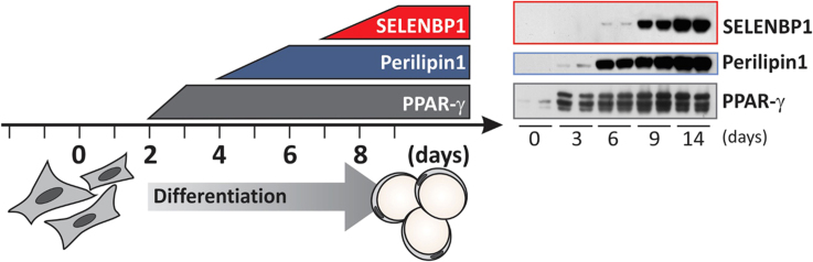
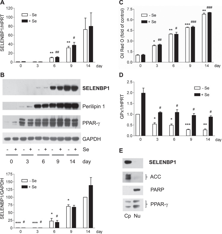
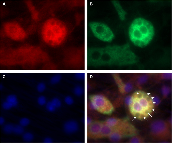
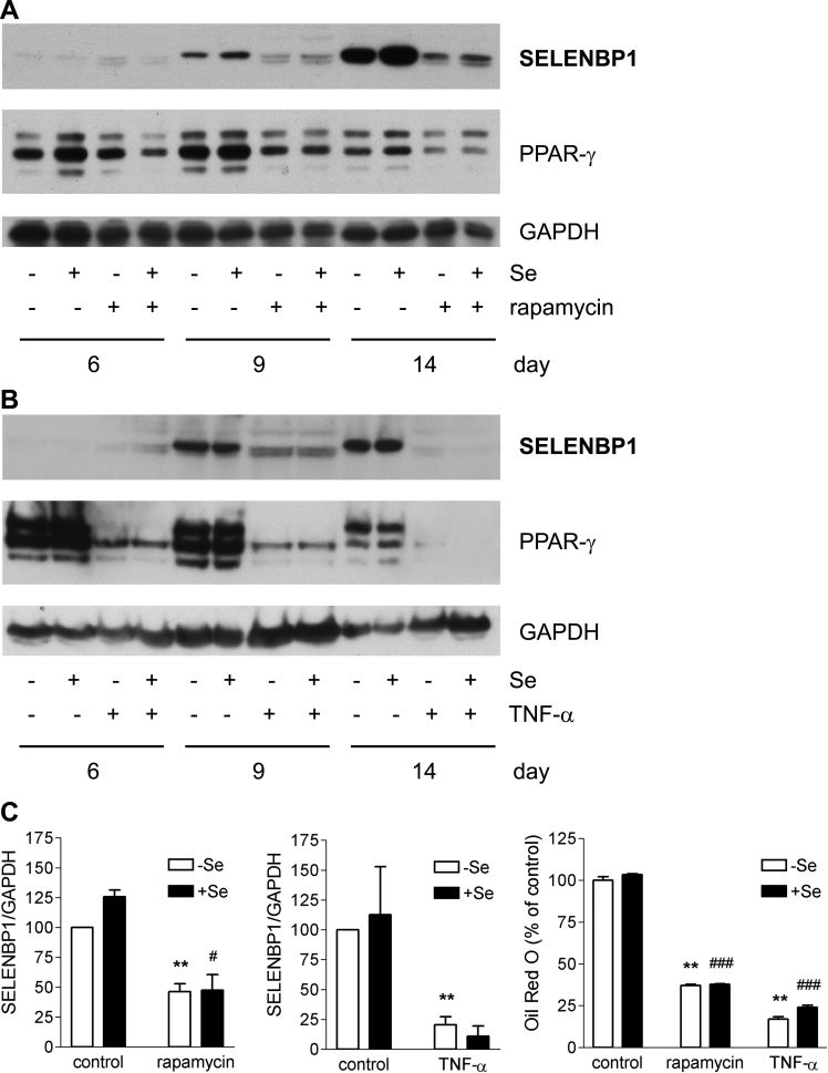
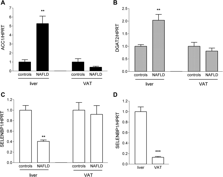
Selenium-binding protein 1 (SELENBP1) has recently been reported to catalyse the oxidation of methanethiol, an organosulfur compound produced by gut microbiota. Two of the reaction products of methanethiol oxidation, hydrogen peroxide and hydrogen sulphide, serve as signalling molecules for cell differentiation. Indeed, colonocyte differentiation has been found to be associated with SELENBP1 induction. Here, we show that SELENBP1 is induced when 3T3-L1 preadipocytes undergo terminal differentiation and maturation to adipocytes. SELENBP1 induction succeeded the up-regulation of known marker proteins of white adipocytes and the intracellular accumulation of lipids. Immunofluorescence microscopy revealed predominant cytoplasmic localisation of SELENBP1 in 3T3-L1 adipocytes, as demonstrated by co-staining with the key lipogenic enzyme, acetyl-CoA-carboxylase (ACC), located in cytosol. In differentiating 3T3-L1 cells, the mTOR inhibitor rapamycin and the pro-inflammatory cytokine tumour necrosis factor alpha (TNF-α) likewise suppressed SELENBP1 induction, adipocyte differentiation and lipid accumulation. However, lipid accumulation per se is not linked to SELENBP1 induction, as hepatic SELENBP1 was down-regulated in high fructose-fed mice despite increased lipogenesis in the liver and development of non-alcoholic fatty liver disease (NAFLD). In conclusion, SELENBP1 is a marker of cell differentiation/maturation rather than being linked to lipogenesis/lipid accumulation.

Keywords: 3T3-L1; Adipogenesis; Lipid accumulation; NAFLD; Selenium, GPx1.


Figures

fx1
Graphical abstract
Fig. 1
Fig. 1
Induction of SELENBP1 in the course of 3T3-L1 adipocyte differentiation. 3T3-L1 cells, cultured with or without 200 nM selenite, underwent adipocyte differentiation for the indicated times (3–14 d). (A) Gene expression of SELENBP1, as analysed by real-time RT-PCR with normalisation against HPRT (n = 4; means ± S.E.M.; **p < 0.01 vs. day 0 (-Se); #p < 0.05, ##p < 0.01 vs. day 0 (+Se)). (B) Protein levels of SELENBP1, as detected by immunoblotting with GAPDH as loading control. Representative immunoblots (upper panel) and densitometric analyses from three independent experiments (lower panel) are shown (n = 3; means ± S.E.M.; *p < 0.05, ***p < 0.001 vs. day 14 (-Se); #p < 0.05 vs. day 14 (+Se)). Induction of perilipin 1 (marker of lipid accumulation) and PPAR-γ (master regulator of adipocyte differentiation) are shown in additional representative immunoblots. (C) Intracellular lipid accumulation, as measured by Oil Red O staining (n = 3; means ± S.E.M.; *p < 0.05, **p < 0.01, ***p < 0.001 vs. day 0 (-Se); # p < 0.05, ## p < 0.01, ### p < 0.001 vs. day 0 (+Se)).). (D) Gene expression of GPx1, as analysed by real-time RT-PCR with normalisation against HPRT (n = 4; means ± S.E.M.; *p < 0.05, **p < 0.01, ***p < 0.001 vs. day 0 (-Se); #p < 0.05 vs. day 0 (+Se)). (E) Localisation of SELENBP1 in mature adipocytes, as depicted in an immunoblot representative of 3 independent experiments. Subcellular fractions of mature 3T3-L1 adipocytes (day 14) were prepared using the NE-PER kit. For control, the cytoplasmic marker ACC, the nuclear marker poly (ADP-ribose) polymerase (PARP) and the adipocyte transcription factor PPAR-γ are shown. Fractions: Cp (cytoplasm), Nu (nucleus).
Fig. 2
Fig. 2
In mature adipocytes, most SELENBP1 co-localises with ACC, the cytosolic key enzyme for fatty acid biosynthesis. 3T3-L1 cells were subjected to adipocyte differentiation for 6 d and subsequent co-immunostaining. (A) SELENBP1 (red), (B) ACC (green), (C) DNA stained with DAPI (blue), (D) merger. The small white arrows in (D) indicate intracellular lipid droplets. The cells are densely packed, as adipocyte differentiation is induced in postconfluent cells undergoing mitotic clonal expansion before terminal differentiation , , . As previously described , the individual cells show unequal rates of differentiation into adipocytes, resulting in a heterogeneous pattern of ACC expression and lipid droplet formation and size.
Fig. 3
Fig. 3
Anti-adipogenic factors suppress SELENBP1 induction in differentiating 3T3-L1 cells. 3T3-L1 cells were subjected to adipocyte differentiation for 6, 9 or 14 d (as indicated) with or without selenite (200 nM) and with or without addition of rapamycin (5 nM) (A) or TNF-α (10 ng/ml) (B). SELENBP1 was detected by immunoblotting. PPAR-γ and GAPDH served as marker for adipocyte differentiation and as loading control, respectively. Immunoblots representative of 3 independent experiments are shown. (C) Relative SELENBP1 protein levels in comparison to intracellular lipid accumulation in 3T3-L1 cells treated with rapamycin or TNF-α for 14 days during adipocyte differentiation. Densitometric analyses of the immunoblots from the experiments with rapamycin (left panel) and TNF-α (middle panel). Lipid accumulation, as measured by Oil Red O staining (right panel) (n = 3; means ± S.E.M.; **p < 0.01 vs. untreated (control) mature adipocytes (-Se); #p < 0.05, ###p < 0.001 vs. untreated (control) mature adipocytes (+Se)).
Fig. 4
Fig. 4
SELENBP1 gene expression is down-regulated in the liver of mice upon induction of NAFLD by a fructose-enriched diet. Female C57BL/6J mice (n = 6/group) were fed either standard chow and plain water (controls) or standard chow and fructose-enriched water (NAFLD) for 16 weeks. Relative ACC1 (A), DGAT2 (B) and SELENBP1 (C). The mRNA levels in liver and VAT of the animals were determined by real-time RT-PCR, with normalisation against HPRT. Liver and VAT mRNA levels, respectively, of the examined target genes in control mice were set as 1 (means ± S.E.M.; **p < 0.01, NAFLD vs. control mice). (D) Relative SELENBP1 mRNA levels in VAT of control mice were calculated in relation to respective values in liver set as 1 (means ± S.E.M.; ***p < 0.001, VAT vs. liver).

References

    1. Labunskyy V.M., Hatfield D.L., Gladyshev V.N. Selenoproteins: molecular pathways and physiological roles. Physiol. Rev. 2014;94(3):739–777. - PMC - PubMed
    1. Rayman M.P. Selenium and human health. Lancet. 2012;379(9822):1256–1268. - PubMed
    1. Steinbrenner H., Speckmann B., Klotz L.O. Selenoproteins: antioxidant selenoenzymes and beyond. Arch. Biochem. Biophys. 2016;595:113–119. - PubMed
    1. Ansong E., Yang W., Diamond A.M. Molecular cross-talk between members of distinct families of selenium containing proteins. Mol. Nutr. Food Res. 2014;58(1):117–123. - PMC - PubMed
    1. Bansal M.P., Oborn C.J., Danielson K.G., Medina D. Evidence for two selenium-binding proteins distinct from glutathione peroxidase in mouse liver. Carcinogenesis. 1989;10(3):541–546. - PubMed

Publication types