. 2018 Nov 21;19(11):3692.
doi: 10.3390/ijms19113692.

Metformin Inhibits Migration and Invasion by Suppressing ROS Production and COX2 Expression in MDA-MB-231 Breast Cancer Cells

Affiliations

Metformin Inhibits Migration and Invasion by Suppressing ROS Production and COX2 Expression in MDA-MB-231 Breast Cancer Cells

Chandler Schexnayder et al. Int J Mol Sci. .

Abstract

Background: Several mechanisms of action have been proposed to explain the apparent antineoplastic functions of metformin, many of which are observed at high concentrations that may not be reflective of achievable tissue concentrations. We propose that metformin at low concentrations functions to inhibit ROS production and inflammatory signaling in breast cancer, thereby reducing metastasis.

Methods: Using the highly invasive MDA-MB-231 breast carcinoma model, we ascertained the impact of metformin on cell viability by DNA content analysis and fluorescent dye exclusion. Migration and invasion assays were performed using a modified Boyden chamber assay and metastasis was ascertained using the chorioallantoic membrane (CAM) assay. PGE2 production was measured by Enzyme-Linked Immunosorbent Assay (ELISA). COX2 and ICAM1 levels were determined by flow cytometry immunoassay.

Results: Metformin acutely decreased cell viability and caused G2 cell cycle arrest only at high concentrations (10 mM). At 100 µM, however, metformin reduced ICAM1 and COX2 expression, as well as reduced PGE2 production and endogenous mitochondrial ROS production while failing to significantly impact cell viability. Consequently, metformin inhibited migration, invasion in vitro and PGE2-dependent metastasis in CAM assays.

Conclusion: At pharmacologically achievable concentrations, metformin does not drastically impact cell viability, but inhibits inflammatory signaling and metastatic progression in breast cancer cells.

Keywords: COX2; ICAM1; metastasis; metformin; reactive oxygen species.


Conflict of interest statement

The authors declare no conflict of interest.

Figures
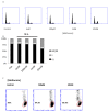
Figure 1
Figure 1
Metformin fails to directly impact breast cancer cell viability at pharmacologically relevant concentrations. (A) MDA-MB-231 breast cancer cells were cultured in the presence or absence of metformin for 72 h. Nuclear DNA content was assessed by propidium iodide staining, and analyzed by flow cytometry. The proportion of cells in each phase is depicted in (B). In (C), cells were cultured in presence or absence of metformin for 48 h, followed by incubation in Sytox Red cell toxicity stain. The dye is excluded from viable cells whose membranes are not compromised (indicated in red). Representative figures are shown. Each assay was performed in quadruplicate. N.S. denotes no significant difference as compared to control.
Figure 2
Figure 2
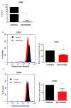
Metformin alleviates endogenous ROS production and subsequent pJNK activation. MDA-MB-231 cells cultured in the presence (red) or absence (black) of metformin (100 µM) for 48 h and subsequently stained with MitoSOX mitochondrial superoxide detection indicator. Fluorescence intensity was ascertained by flow cytometry and was normalized to untreated control. Studies were performed in triplicate, with 10,000 events each, and compared using Student’s t-test, * p ≤ 0.05.
Figure 3
Figure 3
Metformin represses expression of pro-inflammatory markers in breast cancer. (A) MDA-MB-231 cells were incubated with or without metformin for 3 days and levels of PGE2 in the culture supernatant measured by competitive ELISA. MDA-MB-231 breast cancer cells were cultured in the presence or absence of metformin for 48 h after which cells were fixed and immunofluorescently stained for (B) COX2 or (C) ICAM1 protein expression. Staining intensity was measured by flow cytometry and normalized to control for comparison (right of histogram). Flow cytometry assays were performed in quadruplicate with 10,000 events registered per replicate. ELISA was performed with 4 technical repeats on 2 experiments. Significance was determined using Student’s t-test, where * p ≤ 0.05.
Figure 4
Figure 4
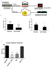
Metformin attenuates breast cancer cell migration, invasion, and metastasis. (A) MDA-MB-231 cells were pre-exposed to metformin for 48 h, collected, and stained with CellTracker Green fluorescent stain. Stained cells were ceded in the upper chamber of a Boyden chamber plate in the absence (B), or the presence (C) of Matrigel coating. The number of transmigratory/invading cells in response to chemoattractant (DMEM with 10% FBS) were enumerated by flow cytometry and normalized to control. (D) Anti-metastatic action of metformin is reversed by PGE2. MDA-MB-231 cells were labeled with CellTracker Green (10 μM) and seeded onto mesh squares placed on the chorioallantoic membrane (CAM) of 10-day old chicken embryos grown ex ovo. After 3 days, portions of the CAM-containing human cells were removed, fixed, stained with DAPI and visualized by fluorescence microscopy using an inverted laser confocal microscope with Z-axis control. Images were taken every 1 μm for approximately 100 μm, and the invasion maximum was defined by the leading edge of fluorescent cell invasion. Significance was determined by ANOVA and Tukey’s post hoc analysis, **** p ≤ 0.01, * p ≤ 0.05.
Figure 5
Figure 5
COX2 expression has prognostic significance in basal, but not Luminal A intrinsic breast carcinoma subtypes. (A) Interrogation of public microarray repositories using KMPLOT online analysis tool (KMPLOT.COM), DMFS was compared between those with high COX2 expression to those with low COX2 expression, in patients with either basal or luminal breast cancer subtypes. The median time of DMFS in the same cohort is summarized in (B).
Figure 6
Figure 6
Model for metformin anti-neoplastic activity. In this model, superoxide is generated in cancer cells during mitochondrial respiration as a by-product of NADH reduction in cancer cells. ROS activity leads to COX2 and ICAM1 expression, as well as induction of other inflammatory mediators, resulting in increased metastatic potential in tumor cells. Metformin disrupts the function of complex I, thereby preventing ROS generation and attenuating metastasis.

References

    1. Col N.F., Ochs L., Springmann V., Aragaki A.K., Chlebowski R.T. Metformin and breast cancer risk: A meta-analysis and critical literature review. Breast Cancer Res. Treat. 2012;135:639–646. doi: 10.1007/s10549-012-2170-x. - DOI - PubMed
    1. Aksoy S., Sendur M.A.N., Altundag K. Demographic and clinico-pathological characteristics in patients with invasive breast cancer receiving metformin. Med. Oncol. 2013;30:590. doi: 10.1007/s12032-013-0590-z. - DOI - PubMed
    1. Xu H., Chen K., Jia X., Tian Y., Dai Y., Li D., Xie J., Tao M., Mao Y. Metformin Use Is Associated with Better Survival of Breast Cancer Patients with Diabetes: A Meta-Analysis. Oncologist. 2015;20:1236–1244. doi: 10.1634/theoncologist.2015-0096. - DOI - PMC - PubMed
    1. Mitsuhashi A., Kiyokawa T., Sato Y., Shozu M. Effects of metformin on endometrial cancer cell growth in vivo: A preoperative prospective trial. Cancer. 2014;120:2986–2995. doi: 10.1002/cncr.28853. - DOI - PubMed
    1. Laskov I., Drudi L., Beauchamp M.-C., Yasmeen A., Ferenczy A., Pollak M., Gotlieb W.H. Anti-diabetic doses of metformin decrease proliferation markers in tumors of patients with endometrial cancer. Gynecol. Oncol. 2014;134:607–614. doi: 10.1016/j.ygyno.2014.06.014. - DOI - PubMed

MeSH terms