

Distribution of Glycerophospholipids in the Adult Human Lens
- PMID: 30469542
- PMCID: PMC6315977
- DOI: 10.3390/biom8040156
Distribution of Glycerophospholipids in the Adult Human Lens
Abstract
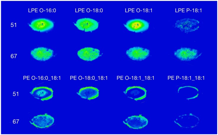
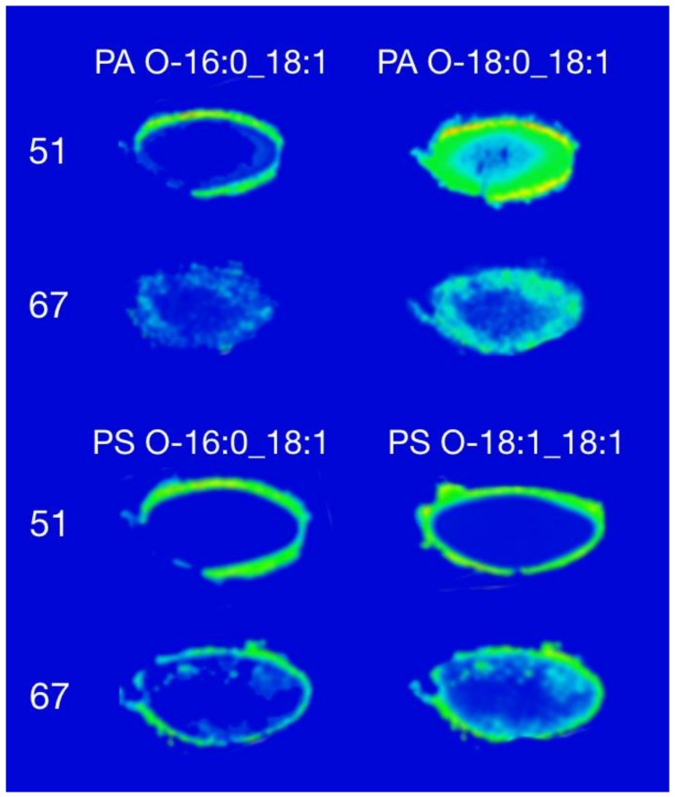
In humans, the age of fibre cells differs across the ocular lens, ranging from those formed before birth in the core of the lens to those formed just prior to death in the outer cortex. The distribution of glycerophospholipids in the adult human lens should reflect this range; however, limited data currently exists to confirm this hypothesis. Accordingly, this study aimed to determine the distribution of glycerophospholipids in adult human lens using mass spectrometry imaging. To achieve this, 20-µm thick slices of two human lenses, aged 51 and 67 were analysed by matrix-assisted laser desorption ionisation imaging mass spectrometry. The data clearly indicate that intact glycerophospholipids such as phosphatidylethanolamine, phosphatidylserine, and phosphatidic acid are mainly present in the outer cortex region, corresponding to the youngest fibre cells, while lyso-phosphatidylethanolamine, likely produced by the degradation of phosphatidylethanolamine, is present in the nucleus (older fibre cells). This study adds further evidence to the relationship between fibre cell age and glycerophospholipid composition.
Keywords: imaging mass spectrometry; lipidomics; phosphatidic acid; phosphatidylserine.
Conflict of interest statement
The authors declare no conflict of interest.
Figures
References
-
- Harding J. Biochemistry of the Eye. Taylor & Francis; Abingdon, UK: 1998.
-
- Heys K.R., Crams S.L., Truscott R.J.W. Massive increase in the stiffness of the human lens nucleus with age: the basis for presbyopia? Mol. Vis. 2004;10:956–963. - PubMed
Publication types
MeSH terms
Substances
LinkOut - more resources
Full Text Sources
